Showing posts with label synaptic turnover. Show all posts

Sunday, July 27, 2025

Hopfield Networks Do Nothing to Explain How a Human Could Remember or Recognize Anything

 Humans have astonishing capabilities for recognizing many different types of things: faces, individual words, quotations, places, musical compositions, and so forth. There is no credible neural explanation for how recognition occurs. There is no robust evidence for any neural correlate of recognition. Brains do not look or act any different when you are recognizing something. For example:

  • The year 2000 study "Dissociating State and Item Components
    of Recognition Memory Using fMRI" found no difference in brain signals of more than 1 part in 100, with almost all of the charted differences being only about 1 part in 500. 
  • The study "Remembrance of Odors Past: Human Olfactory Cortex in Cross-Modal Recognition Memory" found no difference in brain signals of more than 1 part in 200.
  • The study "Neural correlates of auditory recognition under full and divided attention in younger and older adults" found no difference in brain signals of more than 1 part in 500.
  • The study "Neural Correlates of True Memory, False Memory, and Deception" asked people to make a judgment of whether they recognized words, some of which they had been asked to study. The study found no difference in brain signals of more than about 1 part in 300.
  • The study "The Neural Correlates of Recollection: Hippocampal Activation Declines as Episodic Memory Fades" was one in which "participants performed a recognition task at both a short (10-min) and long (1-week) study-test delay." The study found no difference in brain signals of more than about 1 part in 300.
  • The study "The neural correlates of everyday recognition memory" found no difference in brain signals of more than about 1 part in 500.
  • The study "Neural correlates of audio‐visual object recognition: Effects of implicit spatial congruency" was one in which participants attempted a recognition task. The study found no difference in brain signals of more than about 1 part in 200.
Some have claimed that there is something in the brain called a "fusiform face area" that is more active when you are recognizing faces. Such a claim is not well-founded, for reasons I discuss in my post here

But some claim there is some theoretical basis for a little understanding of how a brain could recognize something. For example, the recent paper "Computational models of learning and synaptic plasticity" by neuroscientist Danil Tyulmankov is one of numerous pieces attempting to claim that computer science work provides models shedding insight on how a brain might learn. Such claims are unfounded because of the vast physical differences between what is going on in brains and what goes on inside computers. 

On page 7 of his paper Danil Tyulmankov gives us a typical example of someone trying to trick us into thinking that some computer software technique has some relevance to explaining how a brain could recognize something. Under a heading of "Memory paradigms" and subheadings of "Recall" and "Associative Memory" he states this:

"The colloquial use of 'memory' commonly refers to declarative memory (also called explicit memory) – the storage of facts (semantic memory) or experiences (episodic memory) – which requires intentional conscious recall. One of the most influential models of recall is the associative memory network (Figure 1a), also known as the Hopfield network (Hopfield, 1982). The model’s objective is to store a set of items ... such that when a perturbed version ... of one of the items is presented, the network retrieves the stored item that is most similar to it. For example, given a series of images, as well as a prompt where one of the images is partially obscured, the network would be able to reconstruct the full image. More abstractly, given a series of lived experiences, this may correspond to a verbal prompt to recall a piece of semantic or autobiographical information." 

We have here the typical shenanigans of one of the persons trying to conflate human memory and computer memory, something made rather easy by the fortunate happenstance that the same word ("memory") is used for two completely different things (human memory and computer memory).  Tyulmankov has given us above a paragraph that starts out with a sentence referring to human memory; he then refers to a purely computer software method with no relevance to human memory; and he then ends the paragraph with another sentence referring only to human memory.  It's kind of like someone trying to make you get the impression that the president of the USA is a dog, by having the first sentence of his paragraph referring to dogs, having the second and third sentence of his paragraph referring to the president of the USA, and then having the last sentence of his paragraph again referring to dogs. 

Let me explain some reasons why Hopfield networks do nothing to explain how a human could remember or recognize anything. Hopfield networks are groups of nodes in which each node has a connection to each of the other nodes in the group. The diagram below illustrates a very simple Hopfield network. Each of the circles is called a node. A Hopfield network might have any number of nodes. In the Hopfield network, the different connections between the nodes might have different numerical values called "strengths." 

Hopfield network

Now, if you search on the Internet, you can find various examples of 
programming code that uses Hopfield networks to store and retrieve information. Sometimes while giving such examples, it is claimed that the code has some relevance to explaining how a brain could remember something. We are sometimes told that Hopfield networks have some relevance to the brain, because just as individual neurons in the brain can each be connected to many other neurons because of synaptic connections, each node in a Hopfield network is connected to each node in the network. To play up the similarity, the nodes of a Hopfield network are sometimes called "neurons," even though such a term is profoundly misleading, because of reasons I will explain below. 

There are, however, very strong reasons why Hopfield networks have no relevance to explaining how a brain could remember something. They are listed in my visual above. I will explain each. 

Reason #1: Neurons do not have any capacity for storing some learned piece of information such as an image, a number or a word. 

In a Hopfield network particular nodes of the network may store some item of information. But a neuron does not have any capacity that we know of for storing some item of learned information. No one has ever found an item of learned information by examining a neuron. Very much tissue has been extracted from the brains of living people, and no one ever found in a neuron something like the letter "A" or the word "cat" or the number "1776."  No one has ever found even a single number such as 0 or 1 stored within a neuron. 

Neurons also have no ability to function as binary switches similar to the light switches controlling whether a light is on or off.  A neuron fires at a varying rate, with very much variation from one minute to the next. There is nothing in a neuron that flips between a permanent "off" state and a permanent "on" state.  So even attempts to depict individual neurons as storing a value of 0 or 1 are invalid. Neurons are not like binary switches. 

The page here provides code for a Hopfield network, using the term "neuron" to describe the nodes of the network. It states, "Each neuron in the network represents a binary unit that can have a state of either +1 (active) or −1 (inactive)."  That does not correspond to the physical reality of neurons over any long time scale.  Over the course of a few seconds, a neuron can switch between between being active and inactive. But over a time span such as days, neurons do not switch between some active state and an inactive state. All neurons are electrically active over a time span of 24 hours. So it is not accurate to imagine some situation persisting over a long time in which one neuron corresponds to a 0, and another neuron corresponds to a 1. Neurons fire at a rate between 1 time per second and 200 times per second, and such firing rates vary unpredictably. 

So as simple a storage task as the storage of the word "dog" cannot occur through some method like that imagined above.  The word "DOG" corresponds to the ASCII numbers 68, 79 and 71, and those three digits correspond to the binary sequence 10100111111101100011.  But we can imagine no group of about 20 neurons storing the binary sequence 0100111111101100011 over a long period such as months, because there can be no situation in which some neurons are inactive over a period of months (corresponding to 0) while other neurons are active over months (corresponding to 1).  All neurons are continually active, and neurons do not have any switch-like feature that could enable binary information storage. Plus there's the fact that the brain has no such thing as an ASCII chart allowing a conversion between letters of the English alphabet and decimal numbers. 

Reason #2: Unlike Hopfield networks in computer software, the connections between neurons are noisy and unreliable

Some programming code using Hopfield networks will typically rely on a simple retrieval procedure in which information is extracted across the network with 100% reliability. That does not correspond to the situation in the brain. Almost all connections in the brain require signals passing across chemical synapses. But chemical synapses do not reliably transmit signals across synapses. Scientific papers say that each time a signal is transmitted across a chemical synapse, it is transmitted with a reliability of 50% or less.  A paper states, "Several recent studies have documented the unreliability of central nervous system synapses: typically, a postsynaptic response is produced less than half of the time when a presynaptic nerve impulse arrives at a synapse." Another scientific paper says, "In the cortex, individual synapses seem to be extremely unreliable: the probability of transmitter release in response to a single action potential can be as low as 0.1 or lower." 

What this means is that computer programs using a Hopfield network to retrieve information are not realistically simulating the brain. Were you to modify such programs to realistically simulate the unreliable synaptic transmission in the brain, such programs would no longer be able to achieve their functions of information retrieval or recognition.

Reason #3: A group of neurons is a "fuzzy boundary" thing that does not make a closed network that can be traversed from beginning to end

To understand this reason, let us look at how neurons are arranged in the brain. A typical neuron has very many synapses that connect it to other neurons.  It has been estimated that the brain has about 100 billion neurons, and about 100 trillion synapses. This means the average neuron has about 1000 synapses, each of which is a connection between that neuron and other neurons. All those synapses lock a neuron in place at a particular location, just as the roots of a tree in a dense forest lock that tree into a particular location in the forest.  

The visual below (from the site here) shows some neurons in the brain.  The colors are artificial, supplied to show individual neurons. 


When I search for information on the average distance between neurons, compared to the average size of a neuron, I am told (a) that the average size of the soma at the center of a neuron is about 10-25 micrometers (millions of a meter), and that the average distance between neurons is also about 25 micrometers. So neurons are densely packed in the brain, rather like in the artistic depiction below. 


Now, there is a great problem with any spherical volume of neurons looking like the neurons above.  The problem is that such a volume has no particular spot or neuron that is its beginning, and no particular spot or neuron that is its end. So the volume of neurons cannot be traversed from its beginning to end. For any particular neuron connected to about 1000 other neurons, there is no such thing as a "next neuron" and no such thing as a "previous neuron." 

But a traversal from a beginning to an end is a crucial part of all programming that utilizes Hopfield networks. Traversal from a beginning to an end is crucial to the very idea of a Hopfield network. A Hopfield network does not correspond to a group of neurons, which has a fuzzy boundary and is not like a closed network with a beginning and an end. 

Reason #4: Because of high levels of synaptic remodeling and the short lifetimes of synapse proteins ( < 4 weeks), the strength of connections between neurons rapidly vary randomly.

Hopfield networks include a "weight matrix" that is touted as something similar to the connection between neurons. But in such networks this "weight matrix" is a stable thing. That does not correspond to the connections between neurons, which are ever-varying in a random way. 

Below is a quote from a scientific paper:

"A quantitative value has been attached to the synaptic turnover rate by Stettler et al (2006), who examined the appearance and disappearance of axonal boutons in the intact visual cortex in monkeys.. and found the turnover rate to be 7% per week which would give the average synapse a lifetime of a little over 3 months."

You can read Stettler's paper here2019 paper documents a 16-day examination of synapses, finding "the dataset contained n = 320 stable synapses, n = 163 eliminated synapses and n = 134 formed synapses."  That's about a 33% disappearance rate over a course of 16 days, suggesting an average synapse lifetime of less than three months.
You can google for “synaptic turnover rate” for more information. Synapses typically protrude out of bump-like structures on dendrites called dendritic spines. But those spines have lifetimes of less than 2 years.  Dendritic spines last no more than about a month in the hippocampus, and less than two years in the cortex. This study found that dendritic spines in the hippocampus last for only about 30 days. This study found that dendritic spines in the hippocampus have a turnover of about 40% each 4 days. This 2002 study found that a subgroup of dendritic spines in the cortex of mice brains (the more long-lasting subgroup) have a half-life of only 120 days. A paper on dendritic spines in the neocortex says, "Spines that appear and persist are rare." While a 2009 paper tried to insinuate a link between dendritic spines and memory, its data showed how unstable dendritic spines are.  Speaking of dendritic spines in the cortex, the paper found that "most daily formed spines have an average lifetime of ~1.5 days and a small fraction have an average lifetime of ~1–2 months," and told us that the fraction of dendritic spines lasting for more than a year was less than 1 percent. A 2018 paper has a graph showing a 5-day "survival fraction" of only about 30% for dendritic spines in the cortex.  A 2014 paper found that only 3% of new spines in the cortex persist for more than 22 days. Speaking of dendritic spines, a 2007 paper says, "Most spines that appear in adult animals are transient, and the addition of stable spines and synapses is rare." A 2016 paper found a dendritic spine turnover rate in the neocortex of 4% every 2 days. A 2018 paper found only about 30% of new and existing dendritic spines in the cortex remaining after 16 days (Figure 4 in the paper). 

Furthermore, it is known that the proteins existing between the two knobs of the synapse (the very proteins involved in synapse strengthening) are very short-lived, having average lifetimes of no more than a few days. A graduate student studying memory states it like this:

"It’s long been thought that memories are maintained by the strengthening of synapses, but we know that the proteins involved in that strengthening are very unstable. They turn over on the scale of hours to, at most, a few days."

A scientific paper states the same thing:

Experience-dependent behavioral memories can last a lifetime, whereas even a long-lived protein or mRNA molecule has a half-life of around 24 hrs. Thus, the constituent molecules that subserve the maintenance of a memory will have completely turned over, i.e. have been broken down and resynthesized, over the course of about 1 week.

The paper cited above also states this (page 6):

"The mutually opposing effects of LTP and LTD further add to the eventual disappearance of the memory maintained in the form of synaptic strengths. Successive events of LTP and LTD, occurring in diverse and unrelated contexts, counteract and overwrite each other and will, as time goes by, tend to obliterate old patterns of synaptic weights, covering them with layers of new ones. Once again, we are led to the conclusion that the pattern of synaptic strengths cannot be relied upon to preserve, for instance, childhood memories."

A paper on the lifetime of synapse proteins is the June 2018 paper “Local and global influences on protein turnover in neurons and glia.” The paper starts out by noting that one earlier 2010 study found that the average half-life of brain proteins was about 9 days, and that a 2013 study found that the average half-life of brain proteins was about 5 days. The study then notes in Figure 3 that the average half-life of a synapse protein is only about 5 days, and that all of the main types of brain proteins (such as nucleus, mitochondrion, etc.) have half-lives of 15 days or less.  The 2018 study here precisely measured the lifetimes of more than 3000 brain proteins from all over the brain, and found not a single one with a lifetime of more than 75 days (figure 2 shows the average protein lifetime was only 11 days). 

The paper here states, "Experiments indicate in absence of activity average life times ranging from minutes for immature synapses to two months for mature ones with large weights." The 2025 paper here states that  "the synaptic turnover rate is as high as 1% per day in the visual cortex." That is a rate of about 100% replacement every four months. Quoting an even higher rate of synaptic turnover, the paper here states, "A recent imaging study revealed that the synaptic turnover rate in hippocampal CA1 cells is very high, with an estimated lifetime of 1–2 weeks (Attardo et al., 2015)." 

Similarly, the paper here states that "recent research suggests that dendritic spines in the adult mouse hippocampus are transient with an average lifetime of approximately 10 days," and says, "If this is true, and if turnover is equally likely for all spines, approximately 95-percent of excitatory synapses onto a particular neuron will turn over within 30 days."

When you think about synapses, visualize the edge of a seashore. Just as writing in the sand is a completely unstable way to store information, long-term information cannot be held in synapses. The proteins that make up the synapses are turning over very rapidly (lasting no longer than a few weeks), and the entire synapse is replaced every few months or every several months.  Conversely, humans can reliably remember things they learned or experienced 50 or 60 years ago; and humans can recognize songs, faces, names and quotes that they have not been exposed to in 50 years. For example, lying in bed the other day, there strangely popped into my mind the name "Tobie Tyler." I recognized the name as that of a circus movie involving a boy, one I had not seen or heard mentioned in well over half a century. A Google search confirmed this (I saw the movie around 1960).

Reason #5: There is no ability in the brain to read the strength of the synaptic connections between neurons. 

In a Hopfield network as implemented in computer software code, there is an ability to read the strength of all of the connections between nodes. But the brain has no corresponding ability. The brain has nothing like a synapse strength reader. 

Computer programmers take for granted certain conveniences. Every programmer knows that if he has a data structure named DS, he can run a loop something like the code below, to sum up  the numbers stored in each part of such a data structure:

int nTotal = 0;
int i =0;
for (i = 0; i < DS.length; i++)
    nTotal = nTotal + DS[i];

But while this type of thing is a basic convenience available in the world of programming, it does not correspond to anything possible in the brain. Physically, brains have no way to run loops performing some mathematical or summation operation on each neuron or synapse in a set of neurons or synapses.  A brain cannot sum up the strengths of a set of synapses, nor can a brain even read the exact strength of some particular synapse. Similarly, your muscular system has muscles of various strengths; but there is in your body no such thing as a muscle strength reader; and there never occurs in your body anything like a loop that sums up all the strengths of the muscles in some part of your body. 

In short, while having a small amount of superficial resemblance to the arrangement of neurons and synapses, Hopfield networks and the programing code that use them do not realistically simulate the realities of neurons and synapses.  The ability of Hopfield networks to do certain tasks does nothing to show that the brain is capable of doing such tasks. 

Below is a revealing confession by a neuroscientist named Slotine: "While neuroscience initially inspired key ideas in AI, the last 50 years of neuroscience research have had little influence on the field, and many modern AI algorithms have drifted away from neural analogies." 

Tuesday, July 2, 2024

The Mythology of "Memory Maintenance Molecules"

 In an article at the Nautilus web site, scientist Ken Richardson suggests that his fellow scientists have been guilty of some molecular mythology. He points out that scientists have repeatedly used “action verbs” in describing DNA, telling us that inside DNA are genes that “act,” “behave,” “direct,” “control,” “design,” are “responsible for,” and so forth. But then Richardson tells us “a counter-narrative is building” to correct such erroneous ideas, and then gives us reasons for thinking that genes are merely passive chemical units that do no such things.

Another example of molecule mythology involves a protein called PKMzeta. Some neuroscientists have suggested that PKMzeta has the ability to make memories last for decades in synapses, even though the proteins that make up synapses are very short-lived (having an average lifetime of two weeks or less). Quite a few of the papers or posts spreading this idea were written or co-written by the same person, Todd C. Sacktor. It is never explained clearly by any such theorists how a protein molecule could perform this great feat of magic. For anyone to explain such a thing clearly, he would first need to have a clear theory of how conceptual memories and episodic memories could be stored in synapses. No neuroscientist has ever presented a clear and explicit theory of any such thing. Neuroscientists merely vaguely tell us that somehow memory storage in a brain occurs through “synapse strengthening,” without presenting any clear theory of how that could be memory storage. 

Of course, if you do not have a clear theory of how memories could be stored (for even a few minutes) in synapses, you cannot possibly have a clear theory as to how some protein molecule such as PKMzeta could possibly cause memories stored in synapses to persist for decades, even though the proteins that make up such synapses are very short-lived, lasting an average of less than two weeks. Trying to defend against the charge that synapses are totally unsuitable for storing memories for decades, because of the short lifetimes of the proteins that make up synapses, a scientific paper states, “As long as PKMZ [PKMzeta] remains active and there is an absence of forces which terminate its activity (such as LTD), it will continue to sustain the biochemical changes at the synapse which serve as the neurobiological basis of memory, allowing the memory to persist for durations far exceeding the turnover of its component molecules.” But how could such a miracle of persistence occur, which would be like a message written in wet sand at the seashore persisting for decades, even though the wet sand was being replaced and written over whenever the tide came in? The science paper does not tell us.

Again, we have the case of an “action verb” inappropriately used to describe a molecule. We are told that PKMzeta has a “sustain” super-power allowing it to preserve fantastically complicated information supposedly stored externally in synapses made up of short-lived molecules that are constantly being replaced. There is nothing in the structure of PKMzeta that should cause us to believe it can do any such thing. No theorist has presented an explicit theory as to how anything like PKMzeta could preserve a memory. Such theorists may sometimes present chemical details to impress us, but such details do not constitute a theory unless a theorist gives explicit examples of precisely how specific memories (such as someone's memory of seeing Paris or someone's memory of details learned about World War I) could be permanently stored with the aid of PKMzeta.  No theorist has done any such thing. 

I suppose that if a PKMzeta molecule were able to cause memories to persist despite rapid protein turnover,  we might imagine it as some kind of "genius" molecule that has thoughts like this:

Oh, my goodness, I see that a memory is starting to degrade because of protein turnover! The memory now states, "Ottawa is the capitol of," which isn't even a full English sentence. Why, I'd better synthesize some new proteins to fill in for those proteins that died,  so there can be a nice complete English sentence. Now, what was that country that Ottawa is the capital of?  

Of course, anything the slightest bit like this is very hard to believe in. It would seem that the most minimal requirements that a molecule would have to fulfill in order to be a "memory maintenance molecule" would be the following:

(1) The molecule would have to somehow know whenever a particular protein molecule (that was part of a memory stored in a synapse) had died or disappeared because of the short lifespans of protein molecules.
(2) The molecule would have to somehow cause a replacement protein of the same type to appear in the same place as the vanished molecule, so that the memory did not degrade. 

The problem is that no one can envision a credible scenario under which a molecule could have either of these powers. To imagine how much of a miracle it would be for memories to persist despite constant protein turnover,  you can imagine a homeowner with ten picnic tables in his backyard, each of which is filled with leaves on which a word or two is written. Imagine these leaves spell out narratives, factual information, and ideas. But the problem is that about one day in three there are winds blowing the leaves off of the tables, and scattering them far away. Also, the leaves don't last longer than a year, because they tend to crumble. Now imagine the homeowner has to keep all this information preserved in the leaves, not just for a few nights but for 50 years. That would be a mountainous job.  An equally mountainous job would have to be done if memories were to be preserved in brains despite constant protein turnover causing proteins to persist an average of less than two weeks, and no one has explained how a molecule could possibly do such a feat.  Since synapses face not only rapid protein turnover inside them but also the problem that synapses don't last for longer than a year or two,  they have the same "double degradation" problem that such a homeowner would have with his information written on leaves. 


In the article here, a PKMzeta enthusiast is asked to explain how PKMzeta could cause memories to persist. The scientist gives a lengthy answer which fails to explain how PKMzeta could do such a thing. He merely says "a cluster of PKMzeta molecules can keep themselves turned on perpetually," and then claims that this supposed ability "is a plausible mechanism for memory persistence," without justifying that claim. This fragmentary theorizing is just hand waving. It has never been demonstrated that any cluster of PKMzeta molecules is capable of storing any information (such as a list of words) for a period as long as a month.  We can imagine hypothetical lab experiments that might try to show such a thing, but they have never been done. The paper here refers to "900 synaptic proteins." PKMzeta is only one of those 900 proteins in synapses, being no more common in synapses than an average synapse protein. You don't solve the "short lifetime of proteins" problem by trying to argue that one in 900 of those proteins might somehow have some stability.  As for the scientist's use of the word "plausible," it has been noted by others that "plausible" is the most abused word in theoretical science discourse, and that scientists often carelessly use the word "plausible" without ever doing anything at all to show a likelihood. 

But the PKMzeta enthusiasts have done a few studies which they claim lends credibility to their claims. I will describe a typical such study. A small number of mice are injected with something that suppresses the PKMzeta molecules in their body (or perhaps they are genetically engineered so that they don't have any PKMzeta). Memory experiments are then done. It is sometimes claimed that such mice perform not as well as normal mice. Such experiments have been hailed as support for the “memory maintenance” claims about PKMzeta.

There are several reasons why such studies do not at all show the claims about PKMzeta are correct. The first is that a result such as I described could never show that PKMzeta can save memories from destruction for years. Whenever memory is tested, it's hard to figure out what the cause is for a discrepancy between two test groups. A difference in a test result might be because (1) PKMzeta is involved in perceiving whatever observation is being tested; (2) or that PKMzeta is involved in memory storage; (3) or that PKMzeta is involved in memory retrieval; (4) or that PKMzeta has something to do with attention or focus or visual perception used in a memory test. A test discrepancy could never tell us which of these things was involved. And if some mice did worse in remembering things without PKMzeta, that might justify the small claim that PKMzeta has something to do with memory, but could never justify the vastly more extravagant claim that PKMzeta is capable of preserving memories for decades.

Another reason why such studies do not at all show the claims about PKMzeta are correct has to do with a general malaise in neuroscience. A general problem in modern neuroscience is the production of papers with marginal results that we cannot trust because of things such as too-small sample sizes and publication bias. Let us imagine that neuroscientists want to prove some idea that fits in with their ideological expectations. A great number of experiments might be done, almost all producing no support for the idea. But perhaps 1 in 20 might produce results marginally supporting the idea, probably because of chance variations in data. Now, today negative results are vastly less likely to get published than positive results. So if 19 researchers get a negative result, conflicting with what neuroscientists hope to get, it could be that 10 of them don't even bother to write up their results as a scientific paper, and that the other 9 do write up a paper but don't get it published (because of the journal bias against negative results). However the one researcher who (by chance) got a positive result will write up his result as a scientific paper. Since it will be a result neuroscientists were hoping to get, he will almost certainly get the result published.

This publication bias is a great problem affecting the reliability of scientific research. Because of it we should follow a precautionary neuroscience rule like this: don't believe something has been established unless the result turns up fairly consistently at a high level of significance, in studies with large sample sizes.

Has this happened in regard to memory experiments involving PKMzeta? Not at all. In 2011 a scientist reported three separate studies showing that inhibiting PKMzeta has no effect on memory if tested between 10 and 15 day after the memory forms.  In 2013 two groups of scientists published results conflicting with claims that PKMzeta might allow memories to persist a long time. One study by a team of scientists used genetically engineered mice that had no PKMzeta. It found that such mice “have no deficits in several hippocampal-dependent learning and memory tasks,” and concluded that PKMzeta is not required for memory or learning. Another study by a different team of scientists found that absence of PKMzeta “does not impair learning and memory in mice.” A 2015 study found that inhibiting PKMzeta has no effect on memory in tests performed 30 days after the memory forms. A 2016 paper also found that that inhibiting PKMzeta has no effect on memory in tests performed 30 days after the memory forms.

Such studies would seem to completely debunk claims that PKMzeta enables memories to persist for decades in synapses despite the short lifetimes in the proteins.

The scientists such as Sacktor who helped to spread the PKMzeta myth have tried to fight back with papers such as this 2016 paper. But in that very paper we see evidence that second-rate science is being used to try to prop up claims about PKMzeta. In Figure 7 the scientists tell us how many mice were used for their experiment involving the memory effects of PKMzeta deprivation. They used only 8 mice per study group. That's way too small a sample size to get a moderately convincing result. It is well known that at least 15 animals per study group should be used to get a moderately convincing result. If you use only 8 animals per study group, there's a very high chance you'll get a false alarm, in which the result is due merely to chance variations rather than a real effect in nature.  In fact, in her post "Why Most Published Neuroscience Studies Are False," neuroscientist Kelly Zalocusky suggests that neuroscientists really should be using 31 animals per study group to get a not-very-strong statistical power of .5, and 60 animals per study group to get a fairly strong statistical power of .8.  Compare these numbers to the 8 animals per study group mentioned in Figure 7 of the Sacktor paper. 

This is the same “too small sample size” problem (discussed here) that plagues very many or most neuroscience experiments involving animals. Neuroscientists have known about this problem for many years, but year after year they continue in their errant ways, foisting upon the public too-small-sample-size studies with low statistical power that don't prove anything because of a high chance of false alarms.

If you look up the PRKCZ gene behind the PKMZeta protein molecule, using this page and this page of the Human Protein Database, you will find no characteristics that seem unusual, and nothing suggesting any superstar status. The pages make no mention of the gene even being used in synapses, telling us that the gene is "mainly localized to the cytosol" and "in addition localized to the plasma membrane."   The very idea of some kind of "superstar protein" or "superstar gene" is contrary to the experience in recent decades of scientists, who have found in general that bodily functions almost always involve the coordinated ballet of very many different genes (typically hundreds of them to accomplish a particular task). 

The 2015 scientific paper here shows that PKMzeta rapidly degrades in synapses. The authors say that therefore a stable amount of PKMzeta "would be difficult to maintain at synapses and store memories over long time scales." The paper tells us “There is growing evidence against a role for PKMzeta in memory.” Figure 9 of the paper also shows that a kind of cousin molecule or "isoform" of PKMzeta (PKC lambda) also quickly degrades, experiencing a 50% loss or degradation every 10 hours. So it seems that there is no truth to the idea of PKMzeta (or PKC lambda) as some magic bullet that allows memories to persist for decades in synapses that are constantly having their proteins replaced.

Where does that leave neuroscientists? It leaves them without a leg to stand on in their claims that memories are stored in brains. Based on everything we know about synapses, there is no reason to believe that synapses are capable of storing a memory for even a month, let alone the 50 years that is how long older humans can remember things. As discussed here and here, equally grave problems prevent scientists from creating any credible account of how memories could be encoded into neural states or how seldom-retrieved facts learned many years ago could be instantaneously recalled from a brain that seems to lack any capability for fast look-ups from exact neural positions. We also know (as discussed here and here) that massive damage can occur to brains (such as surgical removal of half of a brain) while producing little effect on memory, which would seem to be impossible if memories are stored in brains. How long before we realize that human memory cannot be a neural thing, but must be a psychic or spiritual phenomenon?

Some people tell tall tales about the protein CAMKII similar to the tall tales told about PKMZeta. We are sometimes told that some alleged autophosphorlyation of CAMKII can help explain stable memories. Most of the reasons I have cited against PKMZeta also apply with equal strength to CAMKII. At this link we are told an experiment debunked the idea that  autophosphorlyation of CAMKII has a role in memory storage.  The lifetime of a CAMKII molecule is only 30 hours, according to this source. The book here makes this statement:

"In the mid-1980's there was much excitement about the idea that autophosphorlyated CaMKII might serve as a self-perpetuating signal that could subserve permanent memory storage. However, a variety of experimental results generated since then suggests that perpetual activation of CaMKII does not occur with LTP-inducing stimulation or memory storage."

This scientific paper says the following:

"Previous models have suggested that CaMKII functions as a bistable switch that could be the molecular correlate of long-term memory, but experiments have failed to validate these predictions....The CaMKII model system is never bistable at resting calcium concentrations, which suggests that CaMKII activity does not function as the biochemical switch underlying long-term memory."

This recent scientific paper says on page 9, "Overall, the studies reviewed here argue against, but do not completely rule out, a role for persistently self-sustaining CaMKII activity in maintaining" long term memory. Another paper says, "The autophosphorylation of CaMKII, once thought to help maintain long-term changes in synaptic strength, has since been revealed to be rather transient." 

Those who have studied the history of science are familiar with epicycles, a complicated speculation that was introduced into Ptolemy's theory of astronomy, to try to fix cases in which the theory did not match observations. We may say these CaMKII speculations and PKMZeta speculations are epicycles intended to fix the failing synaptic theory of memory storage.  But while the Ptolemaic epicycles were exact speculations, the CaMKII speculations and PKMZeta speculations are very vague, failing to specify any exact theory of memory storage. 

Last week a press release from New York University tried to breath life into the dead horse of the mythology of "memory maintenance molecules." The press release was a glaring example of what constantly occurs these days in university press releases: university PR offices boasting about grand accomplishments that were not actually accomplished. We have the untrue claim that a "new study in the journal Science Advances, conducted by a team of international researchers, has uncovered a biological explanation for long-term memories."  No, the study was just more Questionable Research Practices junk science, a study so poorly designed it is a wonder it got published.  We read this:

"It’s been long-established that neurons store information in memory as the pattern of strong synapses and weak synapses, which determines the connectivity and function of neural networks. However, the molecules in synapses are unstable, continually moving around in the neurons, and wearing out and being replaced in hours to days, thereby raising the question: How, then, can memories be stable for years to decades?"

No, it has not ever been established that " neurons store information in memory as the pattern of strong synapses and weak synapses" : no credible theory of how such a thing could work has ever been advanced, no memory information stored in neurons or synapses has ever been found through microscopic examination, and the instability of synapses (including the short lifetimes of their proteins) is the strongest reason for thinking that it cannot possibly be true that "neurons store information in memory as the pattern of strong synapses and weak synapses." 

The press release makes the groundless claim that the junk science paper it is publicizing shows "that KIBRA is the 'missing link' in long-term memories," referring to a molecule called KIBRA.  The press release makes the groundless claim that "their experiments in the Science Advances paper show that breaking the KIBRA-PKMzeta bond erases old memory."

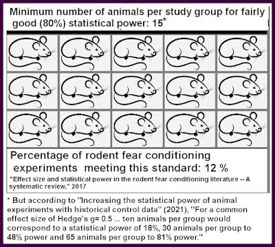
The study is the study "KIBRA anchoring the action of PKMζ maintains the persistence of memory" which you can read here. We have a very bad example of Questionable Research Practices junk science. The study group sizes used are ridiculously small, with study groups as small as only 4 mice and 6 mice, and the average study group size being only about 7 mice. It is frequently pointed out to neuroscientists that experimental studies involving mice are generally worthless unless they use at least 15 subjects per study group; but neuroscientists keep senselessly continuing to use ridiculously low study group sizes.  Why do they do that? Because it allows them to "mine noise," and report false alarms that would vanish if a decent study group size was used. It's rather like someone trying to prove his prophetic powers by publishing a test in which he correctly predicted whether merely four consecutive coin flips were "heads" or "tails," conveniently failing to publish a larger test of his powers involving how well he predicted 15 consecutive coin flips.  You can get all kinds of false alarms when you use tiny sample sizes. 
low statistical power in neuroscience

The junk science paper above relies crucially on an attempt to measure fear, and the attempt to measure fear was the stupid, unreliable technique of attempting to judge "freezing behavior" in mice. All experimental studies relying on estimations of "freezing behavior" are junk science studies, for the reasons I explain in my post here.  There are good reliable ways of measuring fear in rodents, and whether a mouse still has a memory of something the mouse was trained to fear. One good method is to measure heart rate, which dramatically spikes when a rodent is afraid. Another good method is to detect movements in which a mouse avoids a stimulus an animal was trained to fear. The technique is shown in the diagram below:

good way to measure fear in mice

Attempting to measure whether a rodent remembered something fearful by doing estimates of "freezing behavior" is not a reliable way of measuring fear or memory, but instead an unreliable "see whatever you want to see" affair. All studies hinging on so unreliable a method are junk science studies, including the new study by Sacktor mentioned in the New York University press release. Contrary to the claims by Sacktor in his later paper and the claims in the New York University press release, no robust evidence has been produced to show that inhibiting either the KIPRA molecule or the  PKMζ molecule (PKMzeta) does anything to harm the memory of mice. In the paper and the press release Todd Sacktor tells the tall tales of memory maintenance mythology that he likes to tell, unbelievable tales that are not backed up by any robust experiments. 

Genuine "freezing behavior" in an animal would typically only be an instantaneous thing, lasting only a few seconds. The longer the length of time over which "freezing behavior" is judged, the more unreliable such a judgment is as a basis for judging whether the animal recalled a fearful stimulus. In Sacktor's latest paper discussed above, which you can read here,  fear recall is being judged by someone estimating how much an animal was non-moving over a length of four minutes.  That's a particularly unreliable use of the utterly unreliable technique of judging "freezing behavior" as a method of trying to determine whether recall occurred.  

A 2024 scientific paper makes this candid confession, using the phrase "still not completely understood" when it should be saying "are not at all understood":

"Despite over a hundred years of research, the cellular/molecular mechanisms underlying learning and memory are still not completely understood. Many hypotheses have been proposed, but there is no consensus for any of these."

The paper calls Sacktor's speculations about the  PKMζ molecule (PKMzeta) "very controversial." 

I can give you an example that helps show the difficulty of maintaining stable information from unstable components. Let's suppose a shaving creme company wants to publicize its product. It arranges for a line of people to appear outside of the main branch of the New York Public Library, a line in which each person will be displaying a letter made out of shaving creme. The total line of people will spell out the message: "Sale! 10% off on purchases of Barbisol shaving creme, the world's most comfortable shaving creme."  The letters will look like this, but each letter will rest on the outstretched palms of one person.


The problem is that the shaving creme letters will soon decay. How to keep the advertising message running all day?  There could be a system in which each person stands on a numerical position with a number between 1 and 97. If a person sees his shaving creme letter is disintegrating, he then sends a text message to some phone number, saying something like, "I'm leaving -- I'm at position number 12, and my letter is F."  Then some person getting all these text messages can keep sending one of his helpers to each position mentioned in a message, filling in the letter mentioned in the text message.  Through such a system the message might last all day, even though each letter only lasts for less than an hour. Note well some of the requirements here, which include:

(1) A system for representing words by use of symbolic tokens (the English alphabet). 
(2) Some skill for creating these tokens in shaving creme letters (maybe someone who has practiced this skill). 
(3) An addressing system by which each person in the line knows his ordinal position in the line.
(4) A  message system by which components that are about to fail send out a message to some receiving system notifying it to replace their failing token, telling that receiving system of which token to replace, and what the address was of the token to replace. 

No similar system could ever be possible in the human brain. The human brain has no addresses or position numbers or position notation system or coordinate system. Neurons and synapses have no knowledge of the English alphabet, and no capability of writing synapse states or neuron states corresponding to letters of the English alphabet. A protein about to decay in a synapse would never know it was about to decay, and would never be capable of sending some external receiver a "replace me" message. And such a message could never have the address or position coordinates of a synapse protein to be replaced, because tiny components in the brain have neither  addresses nor position coordinates. When you also  consider that synapses are all entangled in 3D space, making them geometrically more difficult to work with than a simple one-dimensional line, you may start to realize how mythical is the notion that stable memories lasting decades could be made from constantly-replaced components with lifetimes of only a few weeks. 

Postscript: A 2018 paper tells us a little about the appalling state of research practices in research involving rodents:

"There is a crisis in pre-clinical biomedical research
involving laboratory animals. Too many papers publish
results which turn out to be irreproducible. One estimate puts the cost at $28 billion being wasted per annum in the United States alone. The causes of this irreproducibility crisis have not
been fully identified. But it has been known for many
years that experiments are often poorly designed, inadequately analysed, and misreported. A survey of
271 papers chosen at random involving rats, mice
and non-human primates showed that 87% did not
report random allocation of experimental subjects to
the treatments and 86% did not report 'blinding' 
when measuring the results. None of the papers gave
any justification for their choice of sample size, and a
substantial number of papers failed even to state the
sex, age or weight of the animal."

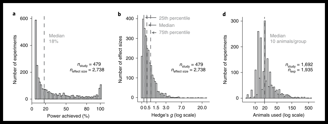
A 2021 paper ("Increasing the statistical power of animal experiments with historical control data" by V. Bonapersona et. al. ) gives us the damning graph below:

poor practices in neuroscience

The graph shows an analysis of 1900+ neuroscience papers. A statistical power of 80% is considered a good goal to reach (when such power is reached there will be about an 80% chance that a reported effect is real). The first graph shows that the average neuroscience paper has a miserably weak statistical power of only about 15%.  The graph on the right shows the number of animals used in these papers, with a median of only about 10 per study group. It is largely because of such low study group sizes that the papers are getting such poor statistical power.  The study group sizes in the Sacktor paper discussed above are sub-standard, even within the dismally poor practices being followed by today's neuroscientists, where the median is a way-too-low number of about 10 animals per study group.  We read this:

"For a common effect size of Hedge’s g= 0.5 (Welch’s independent samples t-test, α=0.05), ten animals per group would correspond to a statistical power of 18%, 30 animals per group to 48% power and 65 animals per group to 81% power...Through a systematic search (Supplementary Notes 1 and 2), we identified a large sample of animal studies in the areas of ‘neuroscience’ and ‘metabolism’ (n...=1,935) that were previously included in meta-analyses (n...=69). These animal studies had an overall median statistical power of 18% (Fig. 1a), which was roughly equal in the two fields (neuroscience, 15%; metabolism, 22%)....We estimated that, at best, 12.5% of a large sample of rodent studies were sufficiently powered (that is, prospective power was larger than 80%). This estimate is a best-case scenario, as it is not yet adjusted for any subsequent multiple testing, experimental bias, P hacking and/or fishing, selective reporting, etc.." 

Postscript: Scientific American has an article covering Sacktor's latest paper, giving us yet another example of its very poor journalism regarding neuroscience research. We have an appalling failure to inform the reader of the most basic facts about Sacktor's latest study. At no point is the reader told that the research merely involved rodents. At no point are we told about the appallingly small study group sizes used, such as only 7 rodents.  Sacktor is allowed to get away with a groundless boast that he "nailed it," and no mention is made of how the study hinged upon an unreliable technique for measuring memory (the faulty judgement of "freezing behavior" discussed above).  The author falls for Sacktor's boasts "hook, line and sinker." 

I may also mention that the study fails to discuss how an effective blinding protocol was followed.  We get a mere sketchy vague  statement that an experimenter judging how much "freezing behavior" occurred was "blind to the conditions," but that does not constitute a description of an effective blinding protocol. When very small study group sizes (such as only seven mice) are used it is easy for a supposedly blind judge to know (by visual recognition) whether or not some animals being analyzed were part of some group that had been chemically treated, and which were desired to be described as acting in a particular way, such as "freezing" more. Effective blinding in an experiment (necessary whenever subjective judgments are made) can only be achieved by following a careful, detailed blinding protocol taking at least a long paragraph to describe; and that apparently did not occur in this study. No reliable measurement or reliable analysis of memory performance has occurred. 

Post-postscript: A perennial promoter of junk neuroscience research, Quanta Magazine had a 2025 article promoting Sacktor's groundless boasts, one with the bogus title "The Molecular Bond That Helps Secure Your Memories." Sacktor's paper with the ridiculously low sample sizes such as only four mice or six mice is passed off as a discovery, with no mention at all of the absurdly low study group sizes used. Another neuroscientist guilty of the same type of very poor research practices (such as the use of way-too-small study group sizes and utterly unreliable "freezing behavior" judgments to try to measure animal recall) is quoted as praising Sacktor's research. Again and again the article uses the word "discovery" or "discovered" for something that was not actually discovered, and again and again the article uses the word "showed" for something that was not actually showed. 

Post-post-postscript: The 2025 paper here states that  "the synaptic turnover rate is as high as 1% per day in the visual cortex." That is a rate of about 100% replacement every four months. If that is the typical rate at which synapses are replaced, synapses cannot be the storage place of memories lasting decades. Quoting an even higher rate of synaptic turnover, the paper here states, "A recent imaging study revealed that the synaptic turnover rate in hippocampal CA1 cells is very high, with an estimated lifetime of 1–2 weeks (Attardo et al., 2015)." 

Thursday, August 5, 2021

Imaging of Dendritic Spines Hint That Brains Are Too Unstable to Store Memories for Decades

Scientists have very fancy equipment for examining brains at very high resolution. But no microscopic examination of a brain has ever proven or even supported the claim that brains store memories.  The most common claim about a brain storage of memories is that memories are stored in synapses. But the paper here confesses, "Very few studies report long-lasting structural changes of synapses induced by behavioral training."

There are two types of ways to examine brain tissue: in vivo or in vitro. An in vitro examination means looking at some tissue that has been removed from an organism, or some tissue in a dead organism. An in vivo examination means examining tissue in a living organism.  When examining human tissue, there are rather severe constrains on what can be seen in vivo. But there are no constraints on in vitro examinations of newly deceased humans, whenever such humans have donated their bodies to medical science.  The brains of quite a few such humans have been minutely examined with the most sophisticated equipment. No one has ever found evidence of a memory stored in a brain. No one has ever read a memory from a dead person. 

There are a number of ways to do in vivo examinations of the brains of living organisms.  One technique is called time-lapse two-photon laser microcopy.  Such technology is not good enough to clearly inspect individual synapses, which are very small. But such microscopy is good enough to show what are called dendritic spines. 

A dendritic spine is a tiny protrusion from one of the dendrites of a neuron. The diagram below shows a neuron in the top half of the diagram. Some dendritic spines are shown in the bottom half of the visual. The bottom half of the visual is a closeup of the red-circled part in the top of the diagram. 

dendritic spine

An individual neuron in the brain may have about a thousand such dendritic spines. The total number of dendritic spines in the brain has been estimated at 100 trillion, which is about a thousand times greater than the number of neurons in the brain.  The total number of synapses in the brain has also been estimated at 100 trillion. A large fraction of synapses are connected to dendritic spines. So by studying how long dendritic spines last, we can tell a good deal about how long synapses last. 

It has been hoped that some relation could be drawn between learning and the formation of new dendritic spines.  But scientists try to insinuate a connection between LTP and learning, and a paper says that "Sorra and Harris measuring three-dimensional reconstructed spines from serial section EM pictures, could not find any significant effect of LTP on morphological properties of spines."

No doubt the first scientists who examined dendritic spines were hoping to see some nice regularity and order, perhaps something that might be some kind of coding system by which dendritic spines might store information.  But dendritic spines show no such regularity. Unlike positions in a DNA molecule (which must be one of only four nucleotide base pair types), dendritic spines can be any of many sizes, shapes or lengths. A length of dendrite and its spines (like the length shown in the bottom half of the visual above) seem to bear no resemblance to encoded information.  The vast majority of new dendritic spines do not last longer than a few months.  

Some unconvincing science papers have attempted to suggest a link between learning and dendritic spines.  Here's what goes on in a typical paper of this type:

(1) Some rodent will be given some learning, such as fear conditioning. 
(2) Various dendritic spines will be examined.
(3) Some newly formed dendritic spines will be declared to be "experience dependent," because they appeared while the learning took place. 

It is easy to explain why such papers use an illegitimate methodology. There are very many billions of dendritic spines in the brain, and they come and go rapidly and randomly. So anyone with a good enough microscope could find some stretch of dendritic spines that increased during learning, just as you could find some stretch of dendritic spines that decreased during learning. There is never any good basis for claiming that some stretch of dendritic spines increased because of some particular type of learning.  Similarly, looking around outside I could find some row of leaves that grew bigger when I was studying something, but there would be zero reason for thinking that such an increase was caused by my learning. 

Some studies compare two different sets of subjects, one that was exposed to learning, and another that was not exposed to learning. The studies may report that the subjects exposed to learning had a greater growth of dendritic spines. This is not at all good evidence that dendritic spines have anything to do with learning. We would expect that if dozens of experiments compared sets of dendritic spines undergoing random fluctuations, that some of them would report (purely by chance) that in some of those sets there was a greater growth of dendritic spines. Similarly, if 100 experimenters tracked the pimples of young teenagers with acne both during the first three months of the school year and during summer vacation, some of the experimenters might report greater numbers of new pimples growing during the first three months of the school year, even though there is no causal connection between learning and the number of pimples a teenager may have on his or her skin. 

By examining the tiny protrusions that are dendritic spines, scientists can get some idea of how stable or unstable these dendritic spines are.  If such spines are very unstable, it is a great problem for any theory that memories are stored in synapses.  Unstable dendritic spines would suggest that synapses are unstable, and are unlikely to be a place where memories could be stored for decades.  Even without studying dendritic spines, we have the strongest reason for believing in the instability of synapses: the fact that proteins in synapses have average lifetimes of only a few weeks. 

Dendritic spines last no more than a few months in the hippocampus, and less than two years in the cortex. This study found that dendritic spines in the hippocampus last for only about 30 days. This study found that dendritic spines in the hippocampus have a turnover of about 40% each 4 days. This study found that dendritic spines in the cortex of mice brains have a half-life of only 120 days. The wikipedia article on dendritic spines says, "Spine number is very variable and spines come and go; in a matter of hours, 10-20% of spines can spontaneously appear or disappear on the pyramidal cells of the cerebral cortex." Referring to in vivo observations of dendritic spines in the mouse hippocampus, the paper here says the authors "measured a spine turnover of ~40% within 4 days."  The 2017 paper here ("Long-term in vivo imaging of experience-dependent synaptic plasticity in adult cortex") found the following regarding dendritic spines in the cortex of rodents:

"About 80% of synapses were detectable for a day or longer; about 60% belonged to the stable pool imaged for at least 8 days. Even this stable pool was found to turn over, with only, 50% of spines surviving for 30 days or longer. Assuming stochastic behaviour, we estimate that the mean lifetime of the stable pool would be on the order of 120 days."

We have no good evidence that any dendritic spines survive for more than  a few years. There is an often-cited paper from the year 2000 with the title "Stably maintained dendritic spines are associated with lifelong memories." The title is misleading, like the title of so many scientific papers.  The paper actually found that "a tiny fraction of daily formed new spines (~0.2% of the total spines) could persist for 3–5 months." So the paper found that only 1 in 500 dendritic spines persist for as long as 5 months.  The paper resorts to some dubious math to try to hypothesize that some dendritic spines may last for years. 

More recent papers have made even more clear the high turnover rate of dendritic spines, and have made it seem less likely that any dendritic spines survive for more than a few years.  The 2015 paper 
"Impermanence of dendritic spines in live adult CA1 hippocampus" states the following, describing a 100% turnover of dendritic spines within six weeks:

"Mathematical modeling revealed that the data best matched kinetic models with a single population of spines of mean lifetime ~1–2 weeks. This implies ~100% turnover in ~2–3 times this interval, a near full erasure of the synaptic connectivity pattern."

The paper here states, "It has been shown that in the hippocampus in vivo, within a month the rate of spine turnover approaches 100% (Attardo et al., 2015; Pfeiffer et al., 2018)." The 2020 paper here states, "Only a tiny fraction of new spines (0.04% of total spines) survive the first few weeks in synaptic circuits and are stably maintained later in life."  The author here is telling us that only 1 in 2500 dendritic spines survive more than a few weeks.  Given such an assertion, we should be very skeptical about the author's insinuation that some very tiny fraction of such spines "are stably maintained." No one has ever observed a dendritic spine lasting for years, and the observations that have been made of dendritic spines give us every reason to assume that dendritic spines do not ever last for more than a few years. 

The same studies that show such short lifetimes for dendritic spines show that while they exist, dendritic spines very rarely maintain the same size and shape.  During their short lifetimes, dendritic spines tend to change very much in size and shape.  One paper says that even among a more stable subset of dendritic spines, "The majority of those (~80%) underwent a fluctuation in head size and neck length of more than 10% (~40% even of more than 30%) within 3 to 4 days."

One 2005 paper ("Development of Long-Term Dendritic Spine Stability in Diverse Regions of Cerebral Cortex") claims to have imaged some mouse dendritic spines in a way so that you could compare how badly some spines decayed after an interval of 18 months. The claim is extremely doubtful, because the paper fails to give any description of how so hard a thing to do was accomplished.  Given the lack of a decent description of how this feat was supposedly accomplished, we should regard the claim with high suspicion. What probably occurred is that some piece of dendrite was examined, and later 18 months later the scientists went looking for some other dendrite with a fairly similar appearance. But we can have no confidence that the exact same little stretch of dendrite was found. Because little stretches of dendrites with a few dendritic spines look so similar, it would be very easy for someone to do a "closest appearance" search, and find a little stretch of dendrite that was not the same little stretch of dendrite examined 18 months earlier. We can have no confidence that the two pairs of dendrites shown in Figure 4 of the paper are actually the same little stretch of dendrite in the same organism. From the author's way-too-scanty description of what was going on, we don't even know whether the images are photos from living mice (as opposed to slides of extracted tissue that were photographed 18 months apart).  We also don't know whether the authors went looking for some fairly stable dendritic spines, in a cherry-picking affair, extracting the most stable-looking dendritic spines from some larger pool where very little stability existed.  

We should keep in mind that a typical neuroscientist studying the lifetimes of dendritic spines (a topic also called dendritic spine turnover or dendritic spine remodeling) is someone likely to be trying to show as much stability as he can find. This is because many  neuroscientists eagerly hope (very illogically) that dendritic spines might be something that can help explain the persistence of memory in humans. So when we see some visual showing some dendritic spines persisting over the course of four months, we are probably seeing something that is not common, something that was cherry-picked to show dendritic spines lasting for a relatively long time. Also, neuroscientists are probably overestimating by 50% or more whenever they estimate what fraction of dendritic spines are stable.

The 2022 paper "Stability and dynamics of dendritic spines in macaque
prefrontal cortex" studied  how long  dendritic spines last in a type of monkey. It says, "We found that newly formed spines were more susceptible to elimination, with only 40% persisting over a period of months."  The same study found that "the percentage of elimination for pre-existing spines over 7 days was only 6% on average," which is a rate that would cause complete disappearance of pre-existing dendritic spines within a year. Dealing with a type of monkey, the 2015 paper "In Vivo Two-Photon Imaging of Dendritic Spines in Marmoset Neocortex" tells us that "The loss or gain rate at the 1 d  [one day] interval observed in this study was similar to those in previous studies of layer 5 neurons of the somatosensory cortex of transgenic mice (12% in 3 d [3 days] for both loss and gain; Kim and Nabekura, 2011) and layer 2/3 neurons of ferret V1 by the virus vector method (4% in 1 d [1 day]  for both loss and gain; Yu et al., 2011)."  The reported loss of dendritic spines is a rate that would cause 100% loss within a year. 

Human memories can last a lifetime, but synapses and the dendritic spines they attach to are very unstable "shifting sands" types of things. "Unstable dendritic spines" implies "unstable synapses," which implies that scientists must be wrong when they claim that memories are stored in synapses.  Stable human memories can last for 50 years, so we cannot believe they are stored in things as unstable as synapses and dendritic spines. Studies on the lifetime of the proteins that make up synapses and dendritic spines tell us that such proteins last only a few weeks.  Synapses and dendritic spines are as unstable as fallen maple leaves.  The brain has no place that it could be storing memories that last for decades.

Postscript: The failure of neuroscientists to listen to what dendritic spines are telling us is epitomized by a 2015 review article on dendritic spines, which states, "It is also known that thick spines may persist for a months [sic], while thin spines are very transient, which indicate that perhaps thick spines are more responsible for development and maintenance of long-term memory."  It is as if the writers had forgotten the fact that humans can remember very well  memories that last for 50 years, a length of time a hundred times longer than "months." 

Electron microscope photos of dendritic spines show something that looks nothing like any organized structure capable of storing information. For example:

dendritic spine closeup

Dendritic spines seem to pop up out of dendrites as randomly as twigs pop up from the branches of trees. No one has been able to detect any kind of regularity in the appearance of dendritic spines that might be a  scheme for storing encoded information, just as no one has been able to find any such scheme in the twigs on trees. Just as there does not exist in nature any such thing as a twig reader that scans branches trying to extract encoded information from the twigs of branches, there does not exist anything in the brain that might be a dendritic spine reader. 

2019 paper documents a 16-day examination of synapses, finding "the dataset contained n = 320 stable synapses, n = 163 eliminated synapses and n = 134 formed synapses."  That's about a 33% disappearance rate over a course of 16 days. The same paper refers to another paper that "reported rates of [dendritic] spine eliminations in the order of 40% over an observation period of 4 days."  paper studying the lifetimes of dendritic spines in the cortex states, "Under our experimental conditions, most spines that appear survive for at most a few days. Spines that appear and persist are rare." The rare persistence referred to was only a persistence of a few months. 

The 2023 paper here gives the graph below showing the decay rate of the volume of dendritic spines. It is obvious from the graph that they do not last for years, and mostly do not even last for six months. 


Page 278 of the same paper says, "Two-photon imaging in the Gan and Svoboda labs revealed that spines can be stable over extended periods of time in vivo but also display genesis (generation) and elimination (pruning) at a frequency of 1–4% per week." Something vanishing at a rate of 2% per week will be gone within a year. 

Post-postscript: The 2025 paper here states that  "the synaptic turnover rate is as high as 1% per day in the visual cortex." That is a rate of about 100% replacement every four months. If that is the typical rate at which synapses are replaced, synapses cannot be the storage place of memories lasting decades. Quoting an even higher rate of synaptic turnover, the paper here states, "A recent imaging study revealed that the synaptic turnover rate in hippocampal CA1 cells is very high, with an estimated lifetime of 1–2 weeks (Attardo et al., 2015)."