Showing posts with label dendritic spines. Show all posts

Sunday, August 23, 2026

Groundlessly Boasting Mouse Hibernation Study At Least Reveals the Short Lifetimes of Synapses and Dendritic Spines

 The recent paper "Artificial hibernation reveals synaptic engram architecture associated with memory retention" is another neuroscience paper making a groundless boast in its title. The authors did nothing at all to reveal a "synaptic engram architecture associated with memory retention."  But this weird study involving putting mice into a kind of artificial hibernation does at least reveal something that helps to debunk all claims of synaptic memory storage. 

The final version of the paper is behind a paywall. But when I search for the paper on Google Scholar, it gives me a preprint as the corresponding .pdf file. The preprint is by the same authors, and has an almost identical title, a title of "Artificial hibernation uncovers distinct synaptic engram architecture for memory retention." So I can use that preprint to analyze the research that went on here.  My use of "the paper" below refers to the preprint. 

The paper is one that has many untrue or groundless statements that reflect the author's dogmas and "improperly jumping to conclusions" thinking. Any statements that the authors make using the words "engrams" or "representations" are groundless. These days cognitive neuroscientists are misusing those words very abundantly. Being guilty of "see what you are hoping to see" pareidolia, neuroscientists are frequently claiming without any good warrant to have seen "representations" of this or that in the brain. It's a "Jesus in my toast" kind of affair.  Similarly, whenever neuroscientists use the term "engram" these days, they are making groundless claims in which some cells or synapses are called "engrams," despite a lack of any decent evidence that any such thing as memory storage has occurred in such cells. "Engram" is a word referring to a claimed place of memory storage in the brain. 

The paper is guilty of methodological sins. The study group sizes were ridiculous, consisting of way-too-small study group sizes such as only three mice, only four mice, only five mice, and a study group size imprecisely described as "6-10 mice for each group."  No study like this should be taken seriously as experimental evidence unless it used a study group size of at least 15 or 20 animals per study group. The authors have used the worthless "freezing behavior" method of trying to judge memory recall, which means that what they claim about experimentally demonstrating memory retention should not be regarded as very reliable. Read my post here for why such a method is worthless as a technique for measuring whether a rodent recalled anything. 

But we do get something of value from the paper: some observations of how unstable and short-lived are synapses and dendritic spines. These are observations which help to discredit the popular but groundless dogma that memories are stored in synapses of the brain, which tend to protrude from dendritic spines. 

A dendritic spine is a tiny protrusion from one of the dendrites of a neuron. The diagram below shows a neuron in the top half of the diagram. Some dendritic spines are shown in the bottom half of the visual. The bottom half of the visual is a closeup of the red-circled part in the top of the diagram. 

dendritic spine

Neuroscientists have often speculated that dendritic spines could be some type of memory storage system, although such a speculation is silly. Dendritic spines no more resemble a memory storage system than do the little twigs on the branch of a tree. And while memories can last decades, dendritic spines are very unstable, typically having a lifetime of only weeks or months.  

We read this (the "QIH" refers to a roughly 48-hour period of artificial hibernation produced by the experimenters):

"We also evaluated the similarity of the locations of all dendritic  spines before and after QIH. The similarity in spine locations between Day 1 and 3, with QIH in between, was 0.91 on average, which was not significantly different from the similarity between Day 3 and 7 without QIH (0.88)."

So in a mere two days or four days, the dendritic spines on the mice lost about 9% of their previous appearance. You can extrapolate from that to get a dendritic spine average lifetime of only several weeks, which is consistent with previous observations by others reported here. Because synapses are typically attached to dendritic spines, anything we learn about the short lifetime of dendritic spines is something also telling us about the short lifetime of synapses. 

The paper also tells us something about the lifetime of synapses. It states this, referring to a hibernation period of only two days. 

"In the current study, we used artificial hibernation in mice to induce extreme downscaling of the neuronal activity and dendritic structures in the hippocampus. During this hypothermic and hypometabolic state, hippocampal neurons show a ~70% reduction in firing rate and the elimination of more than 50% of synapses." 

This is a devastating answer for any one believing that memories are stored in synapses. Extrapolating from such a rate of decay, we would guess that synapses have an average lifetime of only a few days or maybe a few weeks. 

It has long been known that synapses are built from proteins that have very short average lifetimes of only a few weeks.   Richard Huganir led a study that was specifically dedicated to trying to find long-lived proteins in synapses. None were found.  The scientific paper found no such thing. Quite to the contrary, the paper found the following:

  • Studying thousands of brain proteins, the study found that virtually all proteins in brains are very short-lived, with half-lives of less than a week.
  • Table 2 of the paper gives specific half-life estimates for the most long-lasting brain proteins, and in this table only 10 out of thousands of brain proteins had half-lives of 10 days or longer.
  • Of the proteins whose half-life is estimated in Table 2, only one of them has a half-life of longer than 30 days, that protein having a half-life of only 32 days.
  • A graph in the paper indicates that none of the synapse proteins had a half-life of more than 35 days.
Below is a graph from the Huganir paper. It shows that the study found that virtually all proteins in synapses are very short-lived.


Below is another graph from the same paper. It shows that the study found that virtually all proteins in synapses are very short-lived.


Judging from these graphs, none of the proteins found had a half-life of longer than 35 days, and only a few had a half-life of more than 14 days.

The 2025 paper here states this:

"We measured the lifetime of the major excitatory synaptic scaffold protein PSD-95 over a range of spatial scales from brain regions to single synapses. PSD-95–HT lifetimes ranged from 11 to 14 days, depending on the brain region (Fig. 2f,g)."

So if synapses are made from proteins with short lifetimes of only a few days or weeks, we should not be surprised that a researcher would find that about 48 hours would produce a 50% reduction in synapses. Most synapses are attached to dendritic spines. So the previous result about the short lifetime of dendritic spines reinforces the conclusion that synapses have short lifetimes. 

The paper here states, "Experiments indicate in absence of activity average life times ranging from minutes for immature synapses to two months for mature ones with large weights."  The paper suggests that even larger synapses last only two months.  A 2025 paper states that  "the synaptic turnover rate is as high as 1% per day in the visual cortex." Such a rate of turnover is equivalent to a synaptic lifetime of only about a year. Another paper  states that in  hippocampal CA1 cells the synapses have an  estimated lifetime of only 1–2 weeks.

The lifespan of synapses is of very great relevance to whether there is any credibility in the claim that synapses store memories. Old humans can remember very well many things they learned and experienced 50 years ago. But if synapses have short lifetimes, they cannot possibly be a storage place of knowledge humans learned 50 years ago,  and synapses cannot be a storage place of memories humans had 50 years ago.  If synapses have short lifetimes, the synaptic theory of memory is untenable. 

The issue that synapses are made of proteins with short lifetimes has been known for decades. So why on Earth do so many neuroscientists keep claiming that memories are stored in synapses? It's simply that they have no other alternative theory of brain-stored memories that is any better.  Nothing in the brain looks anything like a system for storing, preserving or retrieving learned information or experiences an organism had.  

We know that the body does one type of reading of information: the reading of genetic information from DNA. We understand some of the extremely complex molecular machinery that is involved in such reading. The transcription of DNA requires the RNA Polymerase II protein complex described in my post here. It is a very complex component requiring a special arrangement of more 10,000 amino acids.  Other complex components are required for the reading of DNA, which takes minutes. 

If information were to be stored in synapses or dendritic spines, there would have to be very complex components capable of reading such information. But no trace of any such component can be found. If any such component existed, it would require minutes to read information from synapses and dendritic spines.  Such a hypothetical reality would be utterly incapable of explaining the wonder of instant memory recall. If you ask me, "Who was Abraham Lincoln?" it does not take me minutes to give a detailed answer. I will instantly answer: "The US president during the American Civil War who was assassinated in 1865." 

A search for "DNA reading" on Google Scholar gives ten pages of search results, because DNA reading is a real thing. A search for "synapse reading" on Google Scholar produces only one page of six results. One of these results does not refer to synapse reading; another result is in a foreign language; and the other four results do not refer to a reading of synapses in the human body, but merely refer to something going on in computer  systems in which parts of the system are being inappropriately called "synapses."  We get results so weak because "synapse reading" is not a real thing.  A search for "dendritic spine reading" on Google Scholar gives zero results. 

Tuesday, November 4, 2025

Neuroscientists May Peddle Big BS About the Tiniest Bumps

There are a number of ways to do in vivo examinations of the brains of living organisms.  One technique is called time-lapse two-photon laser microcopy.  Such technology is not good enough to clearly inspect individual synapses, which are very small. But such microscopy is good enough to show what are called dendritic spines. 

A dendritic spine is a tiny protrusion from one of the dendrites of a neuron. The diagram below shows a neuron in the top half of the diagram. Some dendritic spines are shown in the bottom half of the visual. The bottom half of the visual is a closeup of the red-circled part in the top of the diagram. 

dendritic spine

An individual neuron in the brain may have about a thousand such dendritic spines. The total number of dendritic spines in the brain has been estimated at 100 trillion, which is about a thousand times greater than the number of neurons in the brain.  The total number of synapses in the brain has also been estimated at 100 trillion. A large fraction of synapses are connected to dendritic spines. So by studying how long dendritic spines last, we can tell a good deal about how long synapses last. 

It has been hoped that some relation could be drawn between learning and the formation of new dendritic spines.  But scientists try to insinuate a connection between LTP and learning, and a paper says that "Sorra and Harris measuring three-dimensional reconstructed spines from serial section EM pictures, could not find any significant effect of LTP on morphological properties of spines."

No doubt the first scientists who examined dendritic spines were hoping to see some nice regularity and order, perhaps something that might be some kind of coding system by which dendritic spines might store information.  But dendritic spines show no such regularity. Unlike positions in a DNA molecule (which must be one of only four nucleotide base pair types), dendritic spines can be any of many sizes, shapes or lengths. A length of dendrite and its spines (like the length shown in the bottom half of the visual above) seem to bear no resemblance to encoded information.  The vast majority of new dendritic spines do not last longer than a few months.  

Some unconvincing science papers have attempted to suggest a link between learning and dendritic spines. The latest is a paper promoted by a freely fantasizing Ohio State University press release that has the fictional title "Dendritic spines: The key to understanding how memories are linked in time."  Everything the press release states in the quote below is flight-of-fancy stuff without any solid basis in fact:

"The study shows that memories are stored in dendritic compartments: When one memory forms, the affected dendrites are primed to capture new information arriving within the next few hours, linking memories formed close in time.

'If you think of a neuron as a computer, dendrites are like tiny computers inside it, each performing its own calculations,' said lead author Megha Sehgal, assistant professor of psychology at The Ohio State University. 'This discovery shows that our brains can link information arriving close in time to the same dendritic location, expanding our understanding of how memories are organized.' "

Sehgal can believe this wild speculation if she wants, but her statement above has no more basis in fact than a claim that the moon  is mostly made of green cheese. The paper that Sehgal is promoting is an example of very low-quality science research. 

The paper is the paper "Compartmentalized dendritic plasticity
in the mouse retrosplenial cortex links contextual memories formed close in time" which you can read here. It is another comedy-of-errors rodent study so badly designed that we should call it a mouse farce. The study hinges upon the totally unreliable "freezing behavior" technique of trying to measure fear recall in rodents. That method is utterly unreliable, for reasons I discuss at length in my post here.  We have the graph below in the paper. You can tell from the number of dots corresponding to each bar that the study group size used was the way-too-small study group size of only 10 mice per study group. 


Every time you see a graph like this, you can be sure the paper is not a credible scientific effort. Trying to judge "freezing behavior" is not a reliable way of measuring recall in rodents. And when a study claims that freezing behavior was produced in rodents after optogenetic stimulation (as this paper does), trying to claim that this was "memory activation," then we have the most unreliable way in which alleged "freezing behavior" can be used, because the very act of doing such optogenetic stimulation can itself produce "freezing behavior," even if no memory is recalled. Referring to immobility in rodents, a paper states, "It is possible to induce freezing by activating a variety of brain areas and projections, including the hippocampus (Liu et al., 2012), lateral, basal and central amygdala (Ciocchi et al., 2010; Johansen et al., 2010; Gore et al., 2015a), periaqueductal gray (Tovote et al., 2016), motor and primary sensory cortices (Kass et al., 2013), prefrontal projections (Rajasethupathy et al., 2015) and retrosplenial cortex (Cowansage et al., 2014)."

Another reason why the paper "Compartmentalized dendritic plasticity in the mouse retrosplenial cortex links contextual memories formed close in time" is a very bad example of low-quality research is its way too-small study group sizes, consisting of study group sizes such as only 4 mice or 9 mice or 10 mice or 12 mice. We have in this paper with 17 authors yet another farce of a study in which the number of authors is greater than the average study group size -- a situation we can very roughly describe using the scornful term "one mouse per scientist" research.  No experimental neuroscience paper involving rodents should be taken seriously if it uses fewer than 15 or 20 subjects per study group; and typically the study group size needed for good statistical power in neuroscience experimental studies is much larger than 20 subjects per study group.  

When experimental scientists have done their job properly, they produce in their papers something called a sample size calculation, in which they calculate the number of subjects needed to produce a result with a good statistical power such as 80%. When bungling  experimental scientists fail to do their job properly, they will make no mention of doing such a calculation, or they will have a statement like this statement we find in the above paper: "No statistical methods were used to predetermine sample sizes but our sample sizes are similar to those reported in previous publications." A statement like this is similar to someone saying, "I didn't pay my taxes the last five years, but my friends also failed to pay their taxes."  It is a very big ongoing disgrace in experimental neuroscience that experimenters are typically failing to use adequate study group sizes. So you do nothing to show that your study used an adequate study group sizes by claiming that similar studies used the study group size you used. 

neuroscience experiments sample sizes

It seems that the paper has failed to even specify the sex of each of the mice that were used (which is quite a failure in a paper relying on freezing behavior judgments, as a tendency to "freeze" when afraid can differ very much between male and female rodents). The paper claims that "the investigator who collected and analyzed the data including behavior, imaging and staining was blinded to the mouse genotypes and treatment conditions." But the paper fails to give a detailed description of any blinding protocol followed, which should cause us to doubt that effective blinding occurred.  A well-designed study of this type would have had a full description of a blinding protocol, to reassure us that the data gathering and analysis was not just a "see what ever you want to see" affair. 

Contrary to the groundless tall tale that Sehgal has told, neither dendrites nor dendritic spines bear the slightest resemblance to any type of calculation device or computing device; and neither dendrites nor dendritic spines bear the slightest resemblance to memory storage devices. No one has ever found a number, token or symbol in a dendrite or dendritic spine, nor has anyone ever found the slightest speck of software in a dendrite or a dendritic spine. Dendritic spines no more resemble calculation devices or computing devices or memory storage devices than do the pimples on the face of a teenager with a bad case of acne. And each dendritic spine is about as short-lived as each such pimple. 

  The 2015 paper "Impermanence of dendritic spines in live adult CA1 hippocampus" states the following, describing a 100% turnover of dendritic spines within about six weeks:

"Mathematical modeling revealed that the data best matched kinetic models with a single population of spines of mean lifetime ~1–2 weeks. This implies ~100% turnover in ~2–3 times this interval, a near full erasure of the synaptic connectivity pattern."

The paper here states, "It has been shown that in the hippocampus in vivo, within a month the rate of spine turnover approaches 100% (Attardo et al., 2015; Pfeiffer et al., 2018)." The 2020 paper here states, "Only a tiny fraction of new spines (0.04% of total spines) survive the first few weeks in synaptic circuits and are stably maintained later in life."  The author here is telling us that only 1 in 2500 dendritic spines survive more than a few weeks. 

The same studies that show such short lifetimes for dendritic spines show that while they exist, dendritic spines very rarely maintain the same size and shape.  During their short lifetimes, dendritic spines tend to change very much in size and shape.  One paper says that even among a more stable subset of dendritic spines, "The majority of those (~80%) underwent a fluctuation in head size and neck length of more than 10% (~40% even of more than 30%) within 3 to 4 days." Units (dendritic spines) so short-lived and unstable cannot be any basis for storing memories that can last so reliably for decades. 

Saturday, April 12, 2025

When PR for Junk Neuroscience Studies Is Passed Off as "Medical News"

The site MedicalXPress.com is a site that positions itself as a medical news site. The site seems to be one of those sites (such as ScienceDaily.com or Eurekalert.com) that mainly just uncritically publishes the latest university and college press releases, and positions its latest collection of these as either "science news" or "medical news." What's wrong with that? Well, for one thing,  university and college press releases these days are  notorious for their hype, exaggerations, misstatements and frequent untrue headlines, when they are announcing newly published research done at their institution.  So if you run a site that mostly publishes unedited the latest latest research press releases of universities and colleges, you are aiding and abetting the proliferation of false and misleading information. 

Another thing wrong with what goes on at MedicalXPress.com is that the site is guilty of frequently trying to pass off very low-quality neuroscience research as "medical news."  The type of junk rodent research that neuroscientists typically do has no relevance to human health, and it is often very misleading to be selling such shoddy work as medical news, particularly since the work did not involve any attempt to develop a medicine or treatment for humans. 

Let's look at an example of some of the news items that have appeared at the site, involving work utterly unworthy of the attention of people interested in keeping up with medical news. A recent article at the site had the untrue headline "Study shows that dendritic plasticity contributes to the integration of memories."  It was an article attempting to persuade us that microscopic structures in brains called dendritic spines have something to do with memory. There is no good evidence that this is true. 

By the third paragraph of the article, we got some baloney shoveling. Scientist Alcino Silva made grandiose boasts about a very low quality study he co-authored, stating, "A few years back, in a landmark study published in Nature in 2016, we demonstrated that memories formed a few hours apart are linked because they are stored in a common set of neurons in the hippocampus." He was referring to his very low-quality junk science study "A shared neural ensemble links distinct contextual memories encoded close in time," which you can read here.  It was a rodent study using way-too-small study group sizes such as only 4 or 7 or 8 mice. No study of this type should be taken seriously unless it uses at least 15 or 20 subjects per study group. The study also hinged upon the utterly unreliable technique of trying to judge recall in rodents by making subjective judgments of so-called "freezing behavior."  All neuroscience studies that use that utterly unreliable technique are examples of junk science, for reasons I explain here.  The fact that this study has been cited over 1000 times shows the dismally dysfunctional state of neuroscience, in which researchers routinely cite very low-quality junk science studies. 

We then have an equally grandiose and equally groundless boast by another neuroscientist. We read, " 'We showed that when mice form two memories close in time, we can see that many of the same somas, dendritic branches, and spines are involved in forming these two memories,' explained Megha Sehgal, the first author and a co-corresponding author of the paper." Nothing of the sort was done. Dendritic spines are constantly forming and disappearing throughout the brains of every mammal. On any day in the brain of any mammal, there are many millions of dendritic spines appearing; there are many millions of dendritic spines disappearing; there are many millions of dendritic spines enlarging; and there are many millions of dendritic spines shrinking. An observation of something happening to dendritic spines is never evidence that those dendritic spines had anything to do with the formation of a memory. 

Later, getting very excited about her grandiose but groundless claims, Seghal states this: "When we forced independent memories to be stored in the same neuronal somas or even the dendrites and found just this simple intervention in one brain region, the retrosplenial cortex, was enough to link these memories!" The claims are groundless. Scientists have no evidence that memories are stored in any place in a brain, and any claim by a scientist that he or she forced a memory to be in some particular place is a bogus boast. 

The groundless boasts being made are based on research reported in the very low-quality paper here, a paper entitled "Compartmentalized dendritic plasticity in the mouse retrosplenial cortex links contextual memories formed close in time." It's the usual type of very low-quality work we see from rodent memory researchers.  The paper uses way-too-small study group sizes such as only 4 mice per group or only 9 mice group or only 12 mice per group. No rodent research paper of this type using fewer than 20 subjects per study group should be taken seriously, unless it mentions that did a sample size calculation showing that fewer than 20 subjects was sufficient to achieve a good statistical power such as 80% (which this paper does not).  Again, we have a paper that hinges upon attempts to measure rodent recall by using the utterly unreliable technique of trying to judge  "freezing behavior." All neuroscience studies that use that utterly unreliable technique are examples of junk science, for reasons I explain here.  

Among the very many reasons why attempting to judge "freezing behavior" is a marker of junk neuroscience is the fact that there is no standard approach as to how such judgments occur, in regard to the interval of time used. So a researcher can try to judge a mouse's immobility for three minutes, and if he gets a claimed "freezing percentage" he likes, he can use that; but if the percentage is not what he likes, he can use only the first two minutes; and if that percentage is not what he likes, he can use only the first minute; and if that percentage is not what he likes, he can use only the first 30 seconds. That is a "see whatever you want to see" kind of deal, rather than trustworthy measurement. To help show that is not going on, a researcher must always list the time interval used for each and every one of his "freezing behavior" graphs, to show that at least the same time interval was used each time. In the case of the the very low quality paper I am discussing here, the paper "Compartmentalized dendritic plasticity in the mouse retrosplenial cortex links contextual memories formed close in time," we see many "freezing behavior" graphs, none of which mention the time interval corresponding to the graph.  This is science at its clumsiest. Were these claimed degrees of freezing behavior occurring over 30 seconds, 60 seconds, 90 seconds, 120 seconds, or 180 seconds? We are not told, and we cannot tell whether the time interval is different for each graph. So we cannot even tell whether there is a match between the technique used and experimental conventions of neuroscientist rodent researchers. 

Because no reliable technique was used to measure fear or recall, we should disbelieve the claim made in the MedicalXPress article that "Silva, Sehgal and their colleagues found that, following their experimental intervention, mice became scared of a box that was previously unimportant to them, simply because the memory of this box was stored in the same dendrites that stored memories of a box in which they experienced an electric shock."  Such researchers could  have used a reliable technique for measuring fear in mice (heart rate measurement), a technique very reliable because heart rate very dramatically spikes when mice are afraid. Like typical rodent memory researchers, the authors chose the unreliable technique of trying to judge "freezing behavior" rather than the reliable method of looking for heart rate spikes in mice. 

We have the usual lame-as-lame-can-be confession in this paper that the authors failed to do a sample size calculation, as good experimental scientists should do. We read, "No statistical methods were used to predetermine sample sizes but our sample sizes are similar to those reported in previous publications." It is a great scandal that neuroscientists routinely use way-too-small group sizes, creating papers without any decent statistical power, papers mostly reporting only false alarms. Trying to excuse yourself by pointing out that other researchers are using the same way-too-small study group sizes is as lame and laughable as saying, "I don't pay my taxes, but lots of my friends also don't pay their taxes." 

neuroscientist confession

The junk science paper "Compartmentalized dendritic plasticity in the mouse retrosplenial cortex links contextual memories formed close in time" is another joke of a neuroscience research paper in which the study group sizes are smaller than the number of authors, which should make us laugh hard and say: "What was the rule here: only one mouse per researcher?"

inadequate sample sizes in neuroscience

You can do a Google image search for "Effect size versus sample size" to get an idea about the relation between the two, and another thing called statistical power. Some of the visuals you will see may be hard to understand. The visual below describes the situation in an easy to understand way. 

neuroscience sample sizes

The smaller the effect size, the larger the study group size needed to show something in a convincing manner (such as a statistical power of 80%).  Most effect sizes in neuroscience are small.  That means in the great majority of cases a study group of 25 or more is needed. No experimental neuroscience should be taken seriously if it uses a smallest study group size smaller than 15 subjects. It is conceivable that a neuroscience experimental study using 15 or 20 subjects might provide modest evidence for something, but only in the very unlikely case of a high effect size. For the much more likely case of an effect size that is only medium or low, then at least about 35 subjects per study group are needed. 

As a general rule, we should be dismissing as junk science any experimental neuroscience study that fails to either use at least 15 subjects per study group or fails to do a sample size calculation to determine whether the number of subjects was adequate to achieve a good statistical power such as 80%. If the smallest study group size is between 15 and 30, we should only regard the paper as being possibly modest evidence for something if the authors did a sample size calculation to show that with the effect size of the type they are dealing with, the study group size they used was adequate to achieve a good statistical power. 

Strangely in this paper with 17 listed authors we have a reference to "The investigator who collected and analyzed the data including behavior, imaging and staining." So there were 17 people listed as paper authors, but only one person "who collected and analyzed the data"? Who was that person? Was it a PhD, or merely a graduate student? Was it someone who had any experience in doing this kind of extremely tricky easy-to-get-wrong work, involving lots of high-tech equipment easy to misuse, and lots of "freezing behavior" estimations so easy to get wrong? Or was it some graduate student fumbling around while doing such work for the first time? We'll never now, because this "investigator who collected and analyzed the data including behavior, imaging and staining" has not been named. The failure of papers such as this to list the specific people who observed things and when they observed the things is another huge reason for distrusting such papers. And why were 17 people listed as authors, when there was only a single "investigator who collected and analyzed the data"?  Elsewhere a scientist tells us, "Anytime you critique a paper in my field, you might think you’re critiquing the senior scientists on the paper, but they usually have a graduate student or a postdoc who wrote the thing." 

What goes on nowadays in science literature is that scientists massively list themselves as authors of papers they did not write, involving research they had no substantial involvement in.  Then scientists make their accomplishments sound ten times greater than they are, by making claims such as "I am the author of 100 scientific papers," when they were merely the co-author of such papers, with the papers typically having a dozen or more listed authors. 

We should distrust or not believe the claim in the paper that "The investigator who collected and analyzed the data including behavior, imaging and staining was blinded to the mouse genotypes and treatment conditions." Blinding is an important feature of well-designed experiments, as it helps to reduce the chance of "see whatever you want to see" kind of bias. Effective blinding requires a well-designed blinding protocol that takes at least a long paragraph to state. Whenever you read a mere one-sentence assertion that some blinding occurred, without any detailed discussion of an effective blinding protocol, the claim that blinding occurred should not be trusted. An effective blinding protocol in a neuroscience experiment typically requires multiple people involved in collecting and analyzing data. For example, one person might perform some intervention (such as injecting something) into one group of mice (not a group of control mice); some other person not knowing which mice got the shot might perform some performance test on both the mice that got the shot and the control mice; and some third person not knowing which mice got the shot might analyze the performance data. But when you have a single "investigator who collected and analyzed the data," there's no way for that person to be blind about which rodents are in the control group -- unless some very complicated and ingenious scheme was followed to assure effective blinding. If there had been so clever a scheme, we may assume that whoever wrote up the paper would want to tell us about such ingenuity. Since no details about a blinding protocol have been given, other than the bare claim that blinding occurred, we should distrust or disbelieve the claim that this single investigator "was blinded to the mouse genotypes and treatment conditions." 

The insinuations in the junk science papers mentioned above (that dendritic spines help store memories) makes no sense. Dendritic spines no more resemble a place of written information than the twigs on trees. And dendritic spines are too unstable to explain memories that can last for decades.  

dendritic spine

 The 2015 paper "Impermanence of dendritic spines in live adult CA1 hippocampus" states the following, describing a 100% turnover of dendritic spines within six weeks:

"Mathematical modeling revealed that the data best matched kinetic models with a single population of spines of mean lifetime ~1–2 weeks. This implies ~100% turnover in ~2–3 times this interval, a near full erasure of the synaptic connectivity pattern."

The paper here states, "It has been shown that in the hippocampus in vivo, within a month the rate of spine turnover approaches 100% (Attardo et al., 2015; Pfeiffer et al., 2018)." The 2020 paper here states, "Only a tiny fraction of new spines (0.04% of total spines) survive the first few weeks in synaptic circuits and are stably maintained later in life."  The author here is telling us that only 1 in 2500 dendritic spines survive more than a few weeks.  Given such an assertion, we should be very skeptical about the author's insinuation that some very tiny fraction of such spines "are stably maintained." No one has ever observed a dendritic spine lasting for years, and the observations that have been made of dendritic spines give us every reason to assume that dendritic spines do not ever last for more than a few years. Conversely, human knowledge and human motor skills can last for 50 years or more, way too long a time to be explained by changes in dendritic spines or synapses, both of which change too much and too frequently to be a stable storage place for human memories. 

The failure of neuroscientists to listen to what dendritic spines are telling us is epitomized by a 2015 review article on dendritic spines, which states, "It is also known that thick spines may persist for a months [sic], while thin spines are very transient, which indicate that perhaps thick spines are more responsible for development and maintenance of long-term memory."  It is as if the writers had forgotten the fact that humans can remember very well  memories that last for 50 years, a length of time a hundred times longer than "months." 

A 2019 paper documents a 16-day examination of synapses, finding "the dataset contained n = 320 stable synapses, n = 163 eliminated synapses and n = 134 formed synapses."  That's about a 33% disappearance rate over a course of 16 days. The same paper refers to another paper that "reported rates of [dendritic] spine eliminations in the order of 40% over an observation period of 4 days." A paper studying the lifetimes of dendritic spines in the cortex states, "Under our experimental conditions, most spines that appear survive for at most a few days. Spines that appear and persist are rare."

The 2023 paper here gives the graph below showing the decay rate of the volume of dendritic spines. It is obvious from the graph that they do not last for years, and mostly do not even last for six months. 


Page 278 of the same paper says, "Two-photon imaging in the Gan and Svoboda labs revealed that spines can be stable over extended periods of time in vivo but also display genesis (generation) and elimination (pruning) at a frequency of 1–4% per week." Something vanishing at a rate of 2% per week will be gone within a year. Discussing the motor cortex, the paper here says, "We found that 3.5% ± 1.2% of spines were eliminated and 4.3% ± 1.3% were formed in motor cortex over 2 weeks (Figures 3J, 3K, and 3O; 224 spines, 2 animals)." An elimination rate of 3.5% over two weeks would result in 90% elimination over a length of one year. 

The 2022 paper "Stability and dynamics of dendritic spines in macaque prefrontal cortex" studied  how long  dendritic spines last in a type of monkey. It says, "We found that newly formed spines were more susceptible to elimination, with only 40% persisting over a period of months." The same study found that "the percentage of elimination for pre-existing spines over 7 days was only 6% on average," which is a rate that would cause complete disappearance of pre-existing dendritic spines within a year. Dealing with a type of monkey, the 2015 paper "In Vivo Two-Photon Imaging of Dendritic Spines in Marmoset Neocortex" tells us that "The loss or gain rate at the 1 d  [one day] interval observed in this study was similar to those in previous studies of layer 5 neurons of the somatosensory cortex of transgenic mice (12% in 3 d [ 3 days] for both loss and gain; Kim
and Nabekura, 2011) and layer 2/3 neurons of ferret V1 by the virus vector method (4% in 1 d [ 1 day] for both loss and gain; Yu et al., 2011)."  The reported loss of dendritic spines is a rate that would cause 100% loss within a year. 

Most synapses are attached to dendritic spines, so all of these findings about the instability and short lifetimes of dendritic spines are also findings about the instability and short lifetimes of synapses. Both synapses and dendritic spines are way, way too unstable to be a credible storage place for human memories that can last for 50+ years. There is no place in the brain that can be reasonably postulated as a storage place allowing memories to persist for 50 years. 

Currently as the main story on MedicalXPress.com is a story groundlessly claiming that researchers found that groups of neurons encode different types of pain. It's another promotion of a junk science paper, because the study group sizes are so small, consisting of study groups such as 6 mice, 5 mice and 4 mice. Neuroscientists these days are guilty of very often making misleading uses of the terms "encode" and "representation," by claiming to have found "encoding" or "representation" when no robust evidence of any such thing was found. 

Thursday, January 16, 2025

Microscopes Will Never Find the Slightest Trace of Learned Information in a Human Brain

A recent story at the LiveScience.com site has the title "Could we ever retrieve memories from a dead person's brain?" The story has many  inaccurate claims, and fails to tell us the most important facts that are relevant to the question being considered. The subtitle of the article makes this claim: "Neuroscientists have identified the physical locations where memories are stored in the brain."  No, they have not done any such thing, and the article fails to present any good evidence that any such thing was done. 

We have this claim about a method to retrieve a memory from a brain:

"With today's technology, retrieving memories might go something like this. First, identify the set of brain cells, or neurons, that encoded a specific memory in the brain and understand how they are connected. Then, activate those neurons to create an approximate neural network, a machine learning algorithm that mimics the way the brain works."

This does not make any sense as an idea about how one would go about trying to start to read a memory from a brain. The first step in such a process would be to use microscopes to look for any speck of a trace of learned information in a brain. No one has ever succeeded in doing any such thing. Microscopic examination of brain tissue has never revealed a single word anyone ever learned. Microscopic examination of brain tissue has never revealed a single letter or character of anything anyone previously learned, and has never revealed a single pixel (an image dot) of anything anyone ever previously saw. Neural networks are a misnamed type of computer technology that do not actually mimic the brain and its physical shortfalls. 

We have quotes by a neuroscientist (Don Arnold) doing some vague hand-waving and speaking as if he knew things he does not actually know. We read,  "Memories are encoded by groups of neurons, Arnold said." That's the kind of vacuous, vague hand-waving that someone may use when he lacks any actual knowledge of how a brain could store memories.  When people make big important-sounding claims that are not well-supported by evidence, they typically speak in a kind of vague, hand-waving way.  For example, when asked where there existed the weapons of mass destruction that the US claimed were in Iraq before invading it in 2003, US defense secretary Donald Rumsfeld said this:

"We know where they are. They're in the area around Tikrit and Baghdad and east, west, south and north somewhat."

Such weapons of mass destruction were never found in Iraq in 2003 or in the next twenty years. 

Reminding me of the Rumsfeld statement, the LiveScience article claims that long-term memories are formed in the hippocampus, a claim not backed up any robust evidence. The main research paper on the hippocampus and memory is the paper "Memory Outcome after Selective Amygdalohippocampectomy: A Study in 140 Patients with Temporal Lobe Epilepsy." That paper gives memory scores for 140 patients who almost all had the hippocampus removed to stop seizures.  Using the term "en bloc" which means "in its entirety" and the term "resected" which means "cut out," the paper states, "The hippocampus and the parahippocampal gyrus were usually resected en bloc."  The "Memory Outcome after Selective Amygdalohippocampectomy" paper does not use the word "amnesia" to describe the results. That paper gives memory scores that merely show only a modest decline in memory performance.  The paper states, "Nonverbal memory performance is slightly impaired preoperatively in both groups, with no apparent worsening attributable to surgery."  In fact, Table 3 of the paper informs us that a lack of any significant change in memory performance after removal of the hippocampus was far more common than a decline in memory performance, and that a substantial number of the patients improved their memory performance after their hippocampus was removed. 

The LiveScience article tells us, "Other parts of the brain store different aspects of a memory, like emotions and other sensory details, according to the Cleveland Clinic." We are referred to a page on some web site of the Cleveland Clinic that has no named author, and is the kind of article that you might get if you asked ChatGPT about how memory works. That Cleveland Clinic page does not actually make the claim that the LiveScience article attributes to it. The Cleveland Clinic does not claim that different aspects of a memory are stored in different places, but merely claims that other parts of a brain "participate in memory processes."

The LiveScience article then makes a false claim, repeating a groundless achievement legend. It states this:

"Neuroscientists have identified engrams in the hippocampuses of mouse brains. For instance, in a 2012 study published in the journal Nature, researchers found the specific brain cells associated with a memory of an experience that induced fear." 

No, there does any exist any robust evidence for engrams (neural storage places of memories) in any animal.  The paper the LiveScience article links to is the 2012 paper “Optogenetic stimulation of a hippocampal engram activates fear memory recall.” That is a very low-quality paper guilty of several bad examples of Questionable Research Practices. We see in Figure 3 of that paper that inadequate sample sizes were used. The number of animals listed in that figure (during different parts of the experiments) are 12, 12, 12, 5, and 6, for an average of 9.4. That is not anything like what would be needed for a moderately convincing result, which would be a minimum of 15 or 20 animals for each study group, and probably more. The experiment relied crucially on judgments of fear produced by manual assessments of freezing behavior, which were not corroborated by any other technique such as heart-rate measurement. All mouse research papers relying on "freezing behavior" judgments are junk-science papers, for reasons I discuss in my post here, "All Papers Relying on Rodent 'Freezing Behavior' Estimations Are Junk Science."  The 2012 study does not describe in detail any effective blinding protocol, which is another bad defect.  The study involved stimulating certain cells in the brains of mice, with something called optogenetic stimulation. The authors have assumed that when mice freeze after stimulation, that this is a sign that they are recalling some fear memory stored in the part of the brain being stimulated. What the authors neglect to tell us is that stimulation of quite a few regions of a rodent brain will produce freezing behavior. So there is actually no reason for assuming that a fear memory was being recalled when the stimulation occurs. 

There does not exist any observational or experimental support for the existence of engrams (memory storage places) in any animal. Some papers have claimed to have produced such evidence, but their claims do not stand up to critical scrutiny. Papers claiming to produce such evidence are generally guilty of multiple types of Questionable Research Practices such as way-too-small study group sizes, lack of a blinding protocol, and the use of one or more unreliable techniques for judging memory performance. 

The LiveScience article then gives us this bit of excuse-making for why no one has ever read a memory from a brain: "The retrieval of a dead person’s memories is further complicated because the discrete parts of a memory are dispersed throughout the brain; for instance sensory details that can also be stored in the parietal lobe and sensory cortex." This is an appeal to the theory that a single memory is stored not in one tiny part of the brain but in multiple scattered parts of the brain. There is no evidence for such a theory, and the theory makes things worse for the person claiming that the brain stores memories, for reasons I discuss in my post "Why the 'A Memory Is Stored Throughout the Brain' Idea Makes Things Much Worse."  If a single memory were to be stored in multiple locations in the brain, then finding all of those locations and assembling them instantly (in a brain without any addresses or indexes or sorting) would be something even more impossible to explain than imagining that the memory existed in a single spot that was instantly found. 

Giving us its second example of claiming a cited source said something that it did not actually say, the LiveScience article states, "Neurons within a given engram are connected through synapses, the spaces between neurons where electrochemical signals travel, according to the National Library of Medicine."  The page that it links to does not ever use the word "engram," and does not refer to either memory or learning. The LiveScience article claims that according to the neuroscientist Arnold, "there is evidence that memories move to different locations as they are consolidated in the brain." There is no robust evidence of any such thing. Neuroscientists have no credible evidence of memories being stored in any part of the brain of any organism, and they do not have any decent evidence of a memory moving around from one part of the brain to another. 

Arnold is quoted as saying, "You get this sort of cascade of neurons that encode these different things, and each one of them is connected in this engram." That is hand-waving. Scientists lack any robust evidence of any such thing as an engram or an encoding of learned information or experiences in the brain. No scientist has a credible detailed theory of how such encoding could occur.  An ocean of difficulties arises when you start to consider the endless problems that would arise when trying to translate human learned knowledge and experiences into brain states through any imaginable system of encoding. Part of the problem is the extreme variety of things that people can learn and experience (concepts, facts, theories, visual  experiences, auditory experiences, smell experiences, taste experiences, pain experiences, touch experiences, and emotional reactions), meaning there could be no simple encoding scheme (something as simple as the genetic code) that could handle even a tenth of all the types of memories people can form. 

We have no discussion of some of the chief facts relevant to the topic discussed. Some of these facts are below:

(1) Human brain tissue has already been exhaustively studied at very high microscopic resolutions. My post "They Stored and Studied Thousands of Brains, But Still Failed to Show Brains Store Memories" discusses how places such as the Lieber Institute have microscopically studied thousands of brains, most of which were preserved very soon after death. The same post describes how Denmark's University of Odense has stored more than 9000 brains, microscopically examining a large fraction of them. Very much healthy brain tissue just-extracted from living patients has been microscopically examined, because normal brain tissue is often extracted from epilepsy patients when operations are done to prevent intractable seizures resistant to medicine. 
(2) Despite all of that microscopic examination, no one has ever found the slightest trace of any learned information by microscopically examining a brain. Microscopic examination of brain tissue has never revealed a single letter or character of anything anyone learned, and has never revealed a single pixel of anything anyone ever saw. It isn't just that no one ever found anything like "The US has 50 states" by microscopically examining brain tissue; it's that no ever found a U or an S from microscopically examining brain tissue. 
(3) The discovery of a bit of learned information from microscopically examining brain tissue is something that would be many times easier to do than the "recreation of a full memory" imagined by the LiveScience article, but neither of these things has occurred. 
(4) Modern microscopic techniques are powerful enough to discover traces of learned information in the brain if they existed, but no such discovery has occurred. 
(5) Nothing in the brain looks like any type of mechanism for storing learned information or storing memories.  Nothing in the brain looks like any type of apparatus for writing learned  information. 
(6) Nothing in the brain looks like any type of mechanism for reading a stored memory. We can imagine how some organism's brain might physically look like something capable of reading information from a particular spot, by means of something like a moving cursor or moving reading component. The brain has no such thing, and the brain has no moving anatomical parts. 
(7) The places claimed to be sites of brain memory storage (synapses) are unstable places of high molecular turnover, where all the proteins last for only a few weeks or less. There is no credible theory of how places so unstable could be storage places for memories that can reliably last for 60 years. 
(8) There is nothing in the brain that looks like learned information stored according to some systematic format that humans understand or do not understand. Even when scientists cannot figure out a code used to store information, they often can detect hallmarks of encoded information. For example, long before Europeans were able to decipher how hieroglyphics worked, they were able to see a repetition of symbolic tokens that persuaded them that some type of coding system was being used. Nothing like that can be seen in the brain. We see zero signs that synapses or dendritic spines are any such things as encoded information. 
(9) Many humans can remember with perfect accuracy very long bodies of text, but synapses in the brain do not reliably transmit information. An individual chemical synapse transmits an action potential with a reliability of only 50% or less, as little as 10%. A recall of long bodies of text would require a traversal of very many chemical synapses. A scientific paper says, "In the cortex, individual synapses seem to be extremely unreliable: the probability of transmitter release in response to a single action potential can be as low as 0.1 or lower."

Below is a diagram from the paper "Materials Advances Through Aberration-Corrected Electron Microscopy." We see that since the time the genetic code was discovered about 1953, microscopes have grown very many times more powerful. The A on the left stands for an angstrom, a tenth of a nanometer (that is, a ten-billionth of a meter). 


Currently the most powerful microscopes can see things about 1 angstrom in width, which is a tenth of a nanometer. How does this compare to the sizes of the smallest units in brains? Those sizes are below:

Width of a neuron body (soma): about 100 microns (micrometers), which is about 1,000,000 angstroms.

Width of a synapse: about 500 nanometers, about 5000 angstroms.  When you search for "width of a synapse," you will commonly get a figure of 20 to 40 nanometers, but that is the width of the synaptic cleft, the gap between two synapses or between a synapse and a dendrite. The full head is much wider, as you can see from the page here.

Width of a dendritic spine: about 50 to 500 nanometers, about 500 to 5000 angstroms.

Length of a dendritic spine: the site here says, "the thin spine neck, which connects the spine to the main dendritic branch, has lengths between 0.04 and 1 μm [microns] and has 'door knob'-shaped head structures with diameters that between 0.5 and 2 μm [microns]." That length dimension is between 400 and 10,000 angstroms; and that head diameter is between 500 and 20,000 angstroms. 

The visual below (from the page here) shows an electron microscope image of a synapse. The width of the synaptic head is more than 500 nanometers (nm). We see nothing that looks like any kind of storage of human learned information. The neurotransmitters inside the spherical vesicles are short-lived chemicals that don't even last a week. 
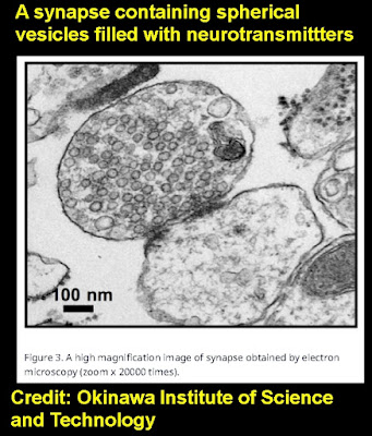
synapse photograph

Below we see a closeup electron microscope photograph of some dendritic spines which are 500 nanometers (5000 angstrom) wide, from the scientific paper here "Ultrastructural comparison of dendritic spine morphology preserved with cryo and chemical fixation," by Tamada et. al.  We see nothing that looks anything like stored learned information or stored memory information. Do a Google search for "diagram of dendritic spine head" and you will get diagrams that look like nothing that could be any system for storing information long-term. The diagrams will show that such dendritic spine heads are just bags of short-lived  proteins and chemicals. A few of these diagrams may show actin filaments looking a bit like a structure, but searching for "lifetime of actin filaments" you will be told that such filaments have lifetimes of only minutes. 

dendritic spine closeup

The only thing in the brain smaller than the structures shown above are protein molecules. But we know a reason why protein molecules cannot be a storage place for memories that can last for 50 years. The reason is that the average lifetime of a brain protein molecule is 1000 times shorter than the longest length that people can remember things.  Proteins in the brain have an average lifetime of two weeks or shorter. 

A scientific paper states this:

"Experience-dependent behavioral memories can last a lifetime, whereas even a long-lived protein or mRNA molecule has a half-life of around 24 hrs. Thus, the constituent molecules that subserve the maintenance of a memory will have completely turned over, i.e. have been broken down and resynthesized, over the course of about 1 week."

Research on the lifetime of synapse proteins is found in the June 2018 paper “Local and global influences on protein turnover in neurons and glia.” The paper starts out by noting that one earlier 2010 study found that the average half-life of brain proteins was about 9 days, and that a 2013 study found that the average half-life of brain proteins was about 5 days. The study then notes in Figure 3 that the average half-life of a synapse protein is only about 5 days, and that all of the main types of brain proteins (such as nucleus, mitochondrion, etc.) have half-lives of 15 days or less.  The 2018 study here precisely measured the lifetimes of more than 3000 brain proteins from all over the brain, and found not a single one with a lifetime of more than 75 days (figure 2 shows the average protein lifetime was only 11 days). 

The paper here states, "Experiments indicate in absence of activity average life times ranging from minutes for immature synapses to two months for mature ones with large weights."

Clearly the resolution of the most powerful microscopes is powerful enough to read memories stored in neurons or synapses or dendritic spines, if such memories existed. And more than 10,000 brains have been microscopically studied in recent years. The failure to microscopically read any  memories from human brain tissue is a major reason for thinking that brains do not store human memories. 

If memories were stored in the brain, roughly about the year 1960 humans would have been able to read learned information stored in brains, when the resolution of microscopes reached about 10 angstroms. If memories were stored in the brain, we would have discovered irrefutable evidence of such a thing about 60 years ago. The total failure to find a single speck of learned information in the brain by microscopic examination is one of the strongest reasons for disbelieving in a brain storage of memories. 

I predict with great confidence that microscopes will never find the slightest trace of learned information in the human brain, because memories are not stored in brains. But we may have in the future some occasional "false alarm" claims to have accomplished such a thing, claims that are not supported by robust evidence. Neuroscientists have often been guilty of both smoke-and-mirrors trickery and pareidolia, when someone claims to see something that isn't really there, typically because he is eagerly scanning large bodies of random, ambiguous data, eagerly hoping to find something that isn't really there. So we may see some pareidolia in which a neuroscientist claims to see a memory by microscopic examination. Such a thing will be like some fervent believer in animal ghosts in the clouds examining thousands of photos of clouds, and claiming that this one or that looks like the shape of an animal. 
evidence-ignoring neuroscientist

Note that in the LiveScience article there is not any mention of any scientist sounding hopeful about a possibility of discovering stored information in the brain. 

This week I was reminded of the ability of the human mind to retain memories for 50 years, contrary to what we would expect from the high molecular turnover in brains.  I had a recollection which proved the ability of the mind to recall very old memories that have not been recalled in half a century. For some reason I recalled a book I had read about 50 years ago, and never since: the science fiction book "Galaxies Like Grains of Sand" by Brian Aldiss. I remembered some lines from the book. I wrote them down on paper like this:

"The mirror of the past lies shattered. The fragments you hold in your hand."

After I wrote this recollection of something I had not read, thought of or heard quoted in fifty years, I borrowed the book on www.archive.org.  I see that the lines were these (almost exactly as I remembered them)

"The long mirror of the past is shattered...Only a few fragments are left, and these you hold in your hand." 

For a person like me, the mirror of the past is not shattered, but remains well preserved after 60 years, contrary to what we would expect if memories were stored in brains with such high molecular turnover and such constant remodeling of synapses and dendritic spines. 

Below is a quote on the same topic from an earlier post discussing why brains cannot be the storage place of very old memories:

"I know for a fact that memories can persist for 50 years, without rehearsal. Recently I was trying to recall all kinds of details from my childhood, and recalled the names of persons I hadn't thought about for decades, as well as a Christmas incident I hadn't thought of for 50 years (I confirmed my recollection by asking my older brother about it). ...Upon looking through a list of old children shows from the 1960's, I saw the title “Lippy the Lion and Hardy Har Har,” which ran from 1962 to 1963 (and was not syndicated in repeats, to the best of my knowledge). I then immediately sung part of the melody of the very catchy theme song, which I hadn't heard in 53 years. I then looked up a clip on a youtube.com, and verified that my recall was exactly correct. I also recently recalled 'The Patty Duke Show' from the 1960's, a show I haven't seen in 50 years, and recalled that in the opening title sequence we saw Patty walking down some stairs. I looked up the title sequence on www.youtube.com, and verified that my 50-year-old memory was correct. This proves that a 53-year-old memory can be instantly recalled."

The prediction I make here is just one of several predictions in my 2019 post "Contrarian Predictions Regarding Biology, the Brain and Technology."  So far my predictions in that post are holding up very well.