A prediction of the dogma that your brain makes your mind is that the more brain injuries you have had, the worse off your mind should be. But a paper in the journal Science ("Effects of Penetrating Brain Injury on Intelligence Test Scores") refers to "the large number of reports describing 'negative' findings -- that is, the absence of demonstrable deficits in test performance, despite the presence of large cerebral lesions, especially in the frontal lobes." The 1957 paper compared IQ tests for 60 armed force members who had their intelligence tested before penetrating brain injuries, and also had their intelligence tested after their brain injuries. Speaking of results on IQ tests, the paper states, "These analyses demonstrated that lesions of the frontal and occipital lobes did not produces a significant decline in score, and that only lesions of parietal or temporal lobes of the left hemisphere showed a significant decrease." The soldiers with lesions in these areas actually performed higher on IQ tests after their penetrating brain injuries, with an average of about a 7% increase:
The only decrease in IQ scores occurred with injuries to the left parietotemporal lobe. These results contradict the results of a new paper entitled "Graph lesion-deficit mapping of fluid intelligence." Instead of finding a decrease in intelligence after right frontal damage as reported by that new paper, the 1957 study found no decrease in intelligence after right frontal damage. The 1957 study used the Army General Classification Test, which is a more reliable test for intelligence than the Raven’s Advanced Progressive Matrices test used by the "Graph lesion-deficit mapping of fluid intelligence" study. One study found less than a 50% correlation between the Raven’s Advanced Progressive Matrices and full-scale IQ. The Raven’s Advanced Progressive Matrices test is a test designed for people of above average intelligence, and is not very suited for testing intelligence damage in people of average intelligence.
There are other reasons for doubting the "Graph lesion-deficit mapping of fluid intelligence" paper. The study hinges upon estimates of "premorbid IQ," someone's IQ before they had some brain damage. The study claims to have something called the "NART IQ," which is an IQ based on a test called the National Adult Reading Test. The National Adult Reading Test can be described as a "quick and dirty" way of very roughly estimating intelligence. It is used by doctors to get a rough idea about a patient's intelligence. Estimates of the correlation between a person's performance on the English NART test and the person's IQ have tended to be about .7, which is a fairly strong correlation, although not a very strong correlation. But a study tested the Dutch version of the NART test and found that it "its current form is not appropriate anymore to estimate premorbid IQ in both young and older adults," having a correlation with intelligence of less than .5.
The study here ("The Relationship of Brain-Tissue Loss Volume and Lesion Location to Cognitive Deficit") tested IQ on 98 veterans with "penetrating brain wounds," finding those with wounds on the right side of the brain to have a mean IQ of 103, and those with wounds on the left side of the brain to have a mean IQ of 99. The paper "Neuropsychological and neurophysiological evaluation of cognitive deficits related to the severity of traumatic brain injury" studied the IQ of 90 patients, dividing them into three categories: mild traumatic brain injury, moderate traumatic brain injury, and severe traumatic brain injury. The mean IQ in each of these groups was about the same, being either 103 or 104. We read that "a surprising finding was that specific intelligence subtests did not show [sensitivity] even for differentiation between severe and mild injury." Such a result is surprising only to those who think your brain makes your mind, not those who reject such an idea.
A recent study was one that attempted to correlate brain volume and intelligence in 262 healthy brain-scanned persons with an age between 55 and 80. An objectionable aspect of the study is that intelligence was measured using only a type of test that young people are known to do better on. We are told, "The Block Design test from the revised form of Wechsler Adult Intelligence Scale [41] was used to assess visuospatial ability and fluid IQ." If we follow the link in that statement, we come to a page telling us, "The results from this test show worse performance in older individuls."
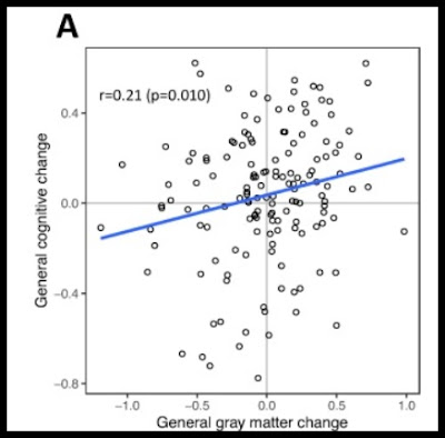
Despite having a chosen a test that is not a good general test of intelligence, presumably to get a more statistically significant result, the authors report only a mild correlation between gray matter change and cognitive change: an R of only .21. The upper left part of their figure 2A (shown below) shows more than 25 cases of people with less gray matter and more intelligence. The result fails to show any clear link between gray matter loss in aging and intelligence.
If the authors had used a better measure of intelligence (the full Wechsler Adult Intelligence test rather than only its Block Design test which seniors do worse on), the authors would have probably got a correlation smaller than the unimpressive correlation of only .21 that they report.
Recently a team of researchers decided to test the brain damage causes memory damage idea by using retirees of the National Football League, people who had played for years in the rough sport known as American football. Although they wear protective helmets, people who have played a long time in the National Football League tend to have had one or more concussions, particularly if they played in positions where concussions more often (such as offensive lineman positions or defensive lineman positions). Described in the press release here, the study "included 53 former NFL players age 50 or older as well as 26 healthy controls and 83 individuals with mild cognitive impairment or dementia who did not play collegiate or professional contact sports and matched as closely as possible to the NFL retirees by age and education." The retired NFL players in the study "had an average of 5.63 concussions, 8.89 years in the NFL, and 115.12 games played."
The press release for the study has a headline of "Head trauma doesn't predict memory problems in NFL retirees, UT Southwestern study shows." We read this:
"Previous studies have reported mixed findings on the relationship between head-injury exposure and neuropsychological functioning later in life. While some investigations have suggested former NFL players may exhibit lower verbal memory and executive function scores, others have not found differences compared to control groups, according to a review of the literature ...The [UT Southwestern] researchers report that retired football players had slightly lower memory scores compared to healthy peer controls but did not find this to be significantly associated with head-injury exposure."
The scientific paper states that except for such slightly lower memory scores "no other group differences were observed, and head-injury exposure did not predict neurocognitive performance at baseline or over time." There was little difference between people who had an average of six concussions and those who had no concussions.
The 2014 paper "No strong evidence for lateralisation of word reading and face recognition deficits following posterior brain injury" has some very good data comparing scores of people with strokes in the rear brain and controls. Table 3 shows no significant difference (denoted as NS) on 16 out of 19 of tests.
A 1996 paper is entitled "Impaired Retrieval From Remote Memory in Patients With Frontal Lobe Damage." There were 7 patients, two of whom had about 50 milliliters of damage (about 5%). Their recognition scores on the Public Events test were only slightly less than normal, with testing covering recognition from 4 decades (Figure 2). The free recall of subjects with frontal lobe damage was a little less than average, and they showed no damage to recognition of Famous Faces (Figure 3), but were a little below average on free recall and cued recall. Figure 4 shows that after an "adjustment" there was basically no difference between the controls and the subjects with frontal lobe damage:


The 4th edition of Hilgard and Atkinson's textbook Introduction to Psychology (1967) made the dubious claim that certain kind of brain lesions can cause the individual to loose their capacity for conceptual thought. They cite a single paper:
ReplyDeleteGoldstein, K., & Scheerer, M. (1941). Abstract and Concrete Behavior: An Experimental Study with Special Tests. Psychological Monographs, 53 [2], whole no. 239.
The book was substantially rewritten in subsequent editions and this paper is no longer referenced in the 7th edition.
This comment has been removed by the author.
DeleteThe paper is behind a paywall, and its abstract does not refer to a brain lesion. An adequate test demonstrating an inability for abstract thought would require something more complicated than a single Sorting Test as described in the abstract. A failure to perform that test well might involve something different, such as a failure to understand spoken instructions. I can imagine various instruction-free tests of abstract thinking. For example, a hungry person could be put in a room with food on a high shelf, reachable only by moving a step ladder to make accessing the food on the shelf. A failure to do that might show an absence of imagination.
DeleteThe paper (which I have not read) is actually a 156 page monograph. I have made it available here:
Deletehttps://drive.proton.me/urls/KNXY5W4EJC#yj99VGQSyMPO
Hilgard and Atkinson only mention a patient who had certain neuromuscular skills such as being able to use a key on a lock or throw a ball into three boxes placed at different distances and yet who was unable to describe these processes or even say which of the boxes was closest. Rather an odd example of an alleged loss of abstract thought (as you wrote, there are many more plausible alternative explanations).