Neuroscientists have often made the claim that the hippocampus is necessary for the formation of new memories. For example, one paper claimed that "clinical evidence indicates that damage to the hippocampus produces anterograde amnesia." Anterograde amnesia is an inability to form new memories. There was never any good evidence for such claims.
To back up claims such as the one above, some people cite the case of patient H.M, a patient with a damaged hippocampus. For example, the paper quoted above states that patient H.M. "became unable to consciously recollect new events in his life or new facts about the world." This is not correct. A 14-year follow-up study of patient H.M. (whose memory problems started in 1953) actually tells us that H.M. was able to form some new memories. The study says this on page 217:
"In February 1968, when shown the head on a Kennedy half-dollar, he said, correctly, that the person portrayed on the coin was President Kennedy. When asked him whether President Kennedy was dead or alive, and he answered, without hesitation, that Kennedy had been assassinated...In a similar way, he recalled various other public events, such as the death of Pope John (soon after the event), and recognized the name of one of the astronauts, but his performance in these respects was quite variable."
Another paper tells us that patient H.M. was able to learn new motor skills., stating this: "H.M. could successfully acquire, and subsequently retain, new motor skills in the context of several other experimental tasks (e.g., rotary pursuit, bimanual tracking, tapping)." Another paper ("Evidence for Semantic Learning in Profound Amnesia: An Investigation With Patient H.M.") states this:
"We used cued recall and forced-choice recognition tasks to investigate whether the patient H.M. had acquired knowledge of people who became famous after the onset of his amnesia. Results revealed that, with first names provided as cues, he was able to recall the corresponding famous last name for 12 of 35 postoperatively famous personalities. This number nearly doubled when semantic cues were added, suggesting that his knowledge of the names was not limited to perceptual information, but was incorporated in a semantic network capable of supporting explicit recall. In forced-choice recognition, H.M. discriminated 87% of postmorbid famous names from foils. Critically, he was able to provide uniquely identifying semantic facts for one-third of these recognized names, describing John Glenn, for example, as 'the first rocketeer' and Lee Harvey Oswald as a man who 'assassinated the president.' Although H.M.’s semantic learning was clearly impaired, the results provide robust, unambiguous evidence that some new semantic learning can be supported by structures beyond the hippocampus proper."
Another paper tells us on page 15 to 16 that patient H. M. was able to draw an accurate map of a bungalow he had moved to after his hippocampus damage, and that he was also able to correctly state the address of that bungalow. On page 20 that paper tells us that patient H.M. had close-to-normal skills in being able to remember visual information:
"In a picture recognition experiment, H.M. was asked to look at complex colorful pictures, for 20 seconds each, and to try to remember them (Freed & Corkin, 1988 et al., 1987). H.M.'s performance revealed a normal recognition of the pictures after 10, 24 hours, 72 hours, and one week after encoding (Augustinack et al., 2014; Corkin, 2020; Freed & Corkin, 1988). Surprisingly, six months later, H.M. could score within one standard deviation from the controls’ average (Freed & Corkin, 1988)."
The myth that patient H.M. lost the ability to form new memories after hippocampus damage is therefore an "old wives' tale" of neuroscience literature, a claim not justified by the facts. I may also note that it is not scientific to cite a patient with one physical issue and some other problem, and to claim or insinuate that the problem was caused by the physical issue. Using the same logic, you could take someone with hair loss and a problem concentrating, and claim that the problem concentrating was caused by the hair loss. Ideas about a cause of something can only be soundly derived from studies involving many patients, not just one or a few.
It is remarkable that scientific papers are still quoting the first paper on patient H.M. as evidence for the claim that the patient could not form new memories. That paper ("LOSS OF RECENT MEMORY AFTER BILATERAL HIPPOCAMPAL LESIONS") failed to document in any scientific way an inability of patient H.M. to form new memories. It provides as its sole evidence an anecdotal account of an interview of April 26, 1955. This is the only evidence provided:
"This was performed on
April 26, 1955. The memory defect was immediately
apparent. The patient gave the date as March, 1953, and
his age as 27. Just before coming into the examining
room he had been talking to Dr. Karl Pribram, yet he
had no recollection of this at all and denied that anyone
had spoken to him. In conversation, he reverted constantly to boyhood events and seemed scarcely to realize
that he had had an operation."
From this scant anecdotal evidence (which easily could involve inaccurate recollection or misinterpretation by one or more doctors), the authors jump to the conclusion that the patient H.M. "appears to have a complete
loss of memory for events subsequent to bilateral medial
temporal-lobe resection 19 months before." This is a conclusion not justified by any scientific tests reported in the paper. In fact, the paper tells us that patient H.M. scored a Memory Quotient score of 67 on the Wechsler Memory Scale (WCS-1) test. That is a score that would have been impossible if patient H.M. had been unable to form new memories. The claim of the authors that patient H.M. "appears to have a complete loss of memory for events subsequent to bilateral medial temporal-lobe resection 19 months before" is a claim contradicted by the test score result reported by the authors in their paper. Judging from the paper
here (particularly Table 1), scoring 67 on the first version of the WCS-1 test would have required getting roughly 40 answers correct on the Wechsler Memory Scale (WCS-1) test, something nobody ever could have done if they could not form new memories.
The main research paper on the hippocampus and memory is the paper "Memory Outcome after Selective Amygdalohippocampectomy: A Study in 140 Patients with Temporal Lobe Epilepsy." That paper gives memory scores for 140 patients who almost all had the hippocampus removed to stop seizures. Using the term "en bloc" which means "in its entirety" and the term "resected" which means "cut out," the paper states, "The hippocampus and the parahippocampal gyrus were usually resected en bloc." The paper refers us to another paper describing the surgeries, and that paper tells us that hippocampectomy (surgical removal of the hippocampus) was performed in almost all of the patients.
The "Memory Outcome after Selective Amygdalohippocampectomy" paper does not use the word "amnesia" to describe the results. That paper gives memory scores that merely show only a modest decline in memory performance. The paper states, "Nonverbal memory performance is slightly impaired preoperatively in both groups, with no apparent worsening attributable to surgery." In fact, Table 3 of the paper informs us that a lack of any significant change in memory performance after removal of the hippocampus was far more common than a decline in memory performance, and that a substantial number of the patients improved their memory performance after their hippocampus was removed.
A 2020 paper is entitled "Preserved visual memory and relational cognition performance in monkeys with selective hippocampal lesions." The paper states this:
"We tested rhesus monkeys on a battery of cognitive tasks including transitive inference, temporal order memory, shape recall, source memory, and image recognition. Contrary to predictions, we observed no robust impairments in memory or relational cognition either within- or between-groups following hippocampal damage."
Citing a previous study, the paper notes that "formation of new memories in the object-in-scene task, one of the most accepted tests of episodic memory used with nonhuman primates, was found to be unaffected by lesions of the hippocampus itself." It also notes that "There is a concerning lack of clear causal evidence for a critical role of the hippocampus in visual memory, episodic memory, recollection, or relational cognition in nonhuman primates."
To test the effects of hippocampus damage, the study authors injected five rhesus monkeys with neurotoxins. They estimate that this damaged about 75% of the hippocampus structures of the monkeys (Figure 1). The monkeys were subjected to a wide variety of cognitive tests. The paper concludes this:
"Contrary to dominant theories, we found no evidence that selective hippocampal damage in rhesus monkeys produced disordered relational cognition or impaired visual memory. Across a substantial battery of cognitive tests, monkeys with hippocampal damage were as accurate as intact monkeys and we found no evidence that the two groups of monkeys solved the tasks in different ways."
These results were similar to those reported by the paper here, entitled "Nonnavigational spatial memory performance is unaffected by hippocampal damage in monkeys." The study tested five monkeys. The study states the following, noting that the monkey that performed the best on one memory test was in the group of hippocampus-damaged monkeys, not the control group of normal monkeys:
"Hippocampal damage did not reduce memory span or slow acquisition. Monkeys with hippocampal damage and control monkeys did not differ in the memory span they achieved during training (mean: HP = 4.4, C = 3.8; median = 4 for both groups; t8 = 1.09, p = .305). The monkey that progressed to the longest memory span (6) was in the hippocampal group (Table 1)."
The 1998 paper "Object Recognition and Location Memory in Monkeys with Excitotoxic Lesions of the Amygdala and Hippocampus" gave 11 monkeys "selective lesions of the amygdala and hippocampus made with the excitotoxin ibotenic acid." According to Table 1, the average hippocampus damage for seven of the monkeys was 73%. We read the following
"Postoperatively, monkeys with the combined amygdala and hippocampal lesions performed as
well as intact controls at every stage of testing. The same
monkeys also were unimpaired relative to controls on an analogous test of spatial memory, delayed nonmatching-tolocation. It is unlikely that unintended sparing of target structures can account for the lack of impairment; there was a
significant positive correlation between the percentage of damage to the hippocampus and scores on portions of the recognition performance test, suggesting that, paradoxically, the
greater the hippocampal damage, the better the recognition."
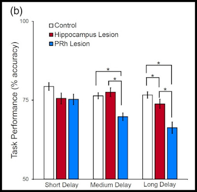
A 2019 paper describing experiments with rhesus monkeys is entitled "Nonnavigational spatial memory performance is unaffected by hippocampal damage in monkeys." A 2023 paper did a meta-analysis of many studies testing memory on monkeys who had been given lesions of the hippocampus. The study was entitled "Reevaluating the role of the hippocampus in memory: A meta-analysis of neurotoxic lesion studies in nonhuman primates." Here is figure 5B from the paper:
The graph is exaggerating the differences, because it using a scale starting at 50% rather than 0%, which is a graph trick that makes small differences look twice as big. Even with the "make the differences look bigger" trick, we see nothing very impressive in regard to the hippocampus. With a short delay and a long delay, there is merely a minimal difference, with the hippocampus-damaged monkeys performing a few percent worse. With a medium delay, the hippocampus-damaged monkeys performed a little bit better. These results fail to back up claims that the hippocampus is crucial for memory.
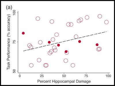
Here is graph 6A from the paper. The graph plots the amount of hippocampus damage on one axis, and the performance on the memory test on the other axis. The graph tells no clear tale. In three of the studies, very good performance (90% or better) occurred despite very high damage of the hippocampus (75% or more ). In seven of the studies, good performance (85% or better) occurred despite 50% or greater hippocampus damage. Figure 3C of the paper shows that the studies involving hippocampus damage of 75% or greater involved about 15 animals per study, while the studies involving hippocampus damage of less than 70% used an average of only about 8 subjects. So we should be granting more weight here to the results shown in the upper right of the diagram below, results showing heavy hippocampus damage and little performance damage.
The 1997 paper "Differential Effects of Early Hippocampal Pathology on Episodic and Semantic Memory" reported on three persons with severe hippocampus damage. We read, "Volumetric measurements derived from
three-dimensional (3D) data sets showed
that in each of the three patients, the hippocampi are abnormally small bilaterally,
with volumes ranging from 43 to 61% of the
mean value of normal individuals (Figs. 2
and 3A)." We learn that "all three patients are
not only competent in speech and language but have learned to read, write, and
spell." We read this:
"With
regard to the acquisition of factual knowledge, which is another hallmark of semantic memory...all three patients obtained
scores within the normal range (Table 2).
A remarkable feature of Beth’s and Jon’s
stores of semantic memories is that they were
accumulated after these patients had incurred the damage to their hippocampi."
In a long footnote to Table 2, we get examples of the three patients answering questions based on quite complex writing in front of them, and answering some common knowledge questions, and the answers sound as good as you or I might give. The authors of this paper attempt to persuade us that the three patients suffered from damage to episodic memory. But they give no very strong evidence of such a thing, mainly mentioning that "none is well oriented in date and time, and
they must frequently be reminded of regularly scheduled appointments and events,
such as particular classes or extracurricular
activities," and that "none can provide
a reliable account of the day’s activities or
reliably remember telephone conversations
or messages, stories, television programs,
visitors, holidays, and so on," leaving us in the dark about what exactly they mean by "reliably." Did they mean 100% correct, 95% correct, or 90% correct? We can't tell. Overall, the paper is inconsistent with claims about the hippocampus being essential for memory.
A scientific paper analyzed the size of the hippocampus in 40 elderly adults. The paper tells us this: "There were no significant correlations between ICV-adjusted hippocampal volumes and age or memory performance (p>.05)."
Postscript: Harvard scientist Karl Lashley did extensive experiments with animals, experiments involving removal or damage to different parts of the brain. In much of what he wrote, it is hard to disentangle the effect of hippocampus damage. But on page 92 of his book Brain mechanisms and intelligence; a quantitative study of injuries to the brain, we have a table that makes it pretty easy to check for how much of an effect damage to the hippocampus has on maze performance in animals who had been trained to run a maze before parts of their brain were removed. The column on the far right lists the type of lesion the animal had. A letter H stands for hippocampus, N stands for No Injury, F stands for Fornix, R stands for right, L stands for left, and the numbers 1, 2 and 3 stand for grade of injury from slight to severe. Lashley states this: "If we select all cases which made more than 25 errors in retention tests, we find that there is no area of [brain] destruction common to all."
These findings were contrary to the dogma that the hippocampus is crucial to memory. Below is what the table tells us about some of the cases. The results are inconsistent with claims that the hippocampus is crucial for memory. The second part of the table can be seen using this link. I have derived the errors per trial by dividing the two rightmost numerical columns in Lashley's table.
|
Case #
|
Total brain tissue loss (%)
|
Hippocampus damage?
|
Total training time, seconds (post-operative)
|
Errors per trial
|
Trials (post-opera-tive)
|
Comment
|
|
98
|
21.1
|
Medium damage on right and left hippocampus
|
310
|
2.2
|
15
|
Much better performance than in case 100, which had no hippocampus damage but similar brain loss damage
|
|
96
|
20.6
|
Medium damage on left hippocampus
|
63 | 5 | 1 | Fair performance, with hippocampus damage and one fifth brain loss
|
|
100
|
21.5
|
None
|
7539
|
10.2
|
75
|
Weak performance, but no hippocampus damage
|
|
107
|
25.4
|
None
|
11536
|
10
|
150
|
Weak performance, but no hippocampus damage
|
|
111
|
28.3
|
Medium damage on right and left hippocampus plus septum damage
|
2230 | 2.6 |
48
|
Much better performance than cases 107 and 100, despite
medium-level hippocampus damage and more brain tissue loss.
|
|
114
|
31.1
|
Small damage on both right and left hippocampus. About one third of brain removed.
|
12
|
1
|
1
|
Very good memory performance despite heavy brain damage and
some hippocampus damage
|
|
116
|
33.9
|
Severe damage on right and left hippocampus
|
2836 | 3.64 |
150
|
Fairly good performance with very bad hippocampus damage and one third
of brain loss, and much better performance than cases 100 and 107 where there
was no hippocampus damage and less brain damage
|
Case 114 and 116 in the data above defy all claims that memories are stored in brains, as they involve good recall performance in animals with one third of the brain removed.
In the paper "Why There Are Complementary Learning Systems in the Hippocampus and Neocortex: Insights From the Successes and Failures of Connectionist Models of Learning and Memory" you can read here, we are told this:
"Some kinds of learning appear to be completely unaffected
by hippocampal system lesions.... Examples of forms of learning that are spared
are gradually acquired skills that emerge over several sessions of
practice, such as the skill of tracing a figure viewed in a mirror
(B. Milner, 1966), reading mirror-reversed print (N. J. Cohen
& Squire, 1980), or anticipating subsequent items in a sequence
governed by a complex stochastic grammar (Cleeremans,
1993). Hippocampal patients also appear to be spared in their
ability to learn the structure common to a set of items: They are
as good as normals in judging whether particular test items
come from the same prototype, or were generated by the same
finite-state grammar, as the members of a previously studied
list (Knowlton, Ramus, & Squire, 1992; Knowlton & Squire,
1993)....In animal studies, it is clear
that some forms of classical or instrumental conditioning of responses to discrete salient cues are unaffected by hippocampal system damage (for reviews, see Barnes, 1988; O'Keefe & Nadel, 1978; Rudy & Sutherland, 1994)."
The paper here refers to humans with hippocampus lesions, and tells us "their acquisition of new skills appears to be completely intact."
The paper "Hippocampal Lesion Patterns in Acute Posterior Cerebral Artery Stroke" did memory tests on patients with lesions of the hippocampus. We have some memory tests on patients who had damage to the hippocampus because of a stroke infarct, who are referred to below as HI patients (hippocampal infarct patients). The groups referred to are those with a right hippocampus infarct, and those with a left hippocampus infarct. We read this:
"In the MMSE, the patients reached a score of 24.30±3.91 (lying in the mildly impaired range), with no difference between groups, t(18)=1.33, P=0.202. In the Clock Drawing Test, the patients reached a score of 2.84±1.26 (at the border of the normal range), with no difference between groups, t(17)=0.51, P=0.618."
In regard to results of a RBMT test of long-term verbal memory, we read this: "Compared to normative samples, the scores of patients with left HI were within the mildly impaired range, whereas the scores of patients with right HI were only slightly below the mean of the normative sample." Overall, this paper supports the claim that the hippocampus is not some crucial component of memory. The people with hippocampus damage have done only slightly less than normal on memory tests.
Post-postscript: One of the main tests used to assess memory in rats is called the Morris Water Maze test. When the test is used, a rat is placed in a tank with a small submerged platform. The water in the tank is so high that the rat is required to swim or tread water to stay afloat, unless it finds the small submerged platform, that is a comfortable place for it rest on, and stay on without swimming or treading water. When the Morris Water Maze test is used a rat is placed in the tank with the submerged platform many different times, and scientists check for how well the rat learns to quickly go to the platform, to get to a comfortable state.
The inventor of the widely used Morris Water Maze test (R. G. M. Morris) tested learning in rats using a Morris Water Maze test. In the 1982 paper here ("Place navigation impaired in rats with hippocampal lesions"), we read of tests involving ten rats with "total hippocampal lesions," 13 rats with "superficial cortical lesions," and 8 control rats that were normal. We are told that "all rats of the hippocampal lesion group were found to have total or near total destruction of the dorsal and ventral hippocampus." So for these rats there was basically total removal of all parts of the hippocampus.
The results are shown below. The rats without hippocampus were able to steadily progress in learning the trick of rapidly going to the submerged platform. Although they learned less quickly than the other two groups of rats, the progress of the rats without any hippocampus was steady, and by about the 30-day mark they had learned the task just as well as the control rats with normal brains. So the rats without hippocampus ended up learning more slowly, but learning just as fully as the rats with normal brains. Clearly it simply is not true that the hippocampus is required to form new memories.
It is thought that the hippocampus may some relation to navigation ability, and the difference shown in the graph above may be purely due to a decrease in navigation ability, without involving any difference in memory storage or memory recall.
The paper "Evidence That the Rat Hippocampus Has Contrasting Roles in Object Recognition Memory and Object Recency Memory" did memory experiments with 16 rats that had "extensive bilateral lesions" of the hippocampus, with a loss of about 80% of the hippocampus. Figure 3 of the paper shows there was almost no difference between the memory performance of the 16 rats with almost no hippocampus and 10 normal rats. We read this:
"There
was no evidence that hippocampal damage impaired object recognition memory even though the rats were tested across retention
intervals (1 min to 120 min) sufficient to capture a full range of
performance levels. This null result is strengthened by the fact that
each rat received many individual trials, far more than given in
conventional spontaneous object recognition memory tasks."
In the paper here an "H-IBO" group of 8 rats received about 90% damage to the hippocampus. This group performed almost as well on memory recognition tests as normal rats (Figure 4).
The 1985 paper here ("THE EFFECTS OF HIPPOCAMPAL LESIONS UPON SPATIAL AND NON-SPATIAL TESTS OF WORKING MEMORY") has a remarkable Figure 5 showing rats that had the "large majority" of the hippocampus removed on both sides of the brain doing better on an object recognition memory test that normal rats. The paper summarizes tests done on rats that had the "large majority" of their hippocampus removed on both sides of the brain:
"The first experiment found no evidence that
extensive hippocampal lesions disrupted either
the acquisition or subsequent performance of a
stimulus recognition task. Additional experiments
helped confirm that the animals had indeed learnt
a non-spatial working memory task and indicated
that the experiment taxed visual recognition
memory. The normal performance of the animals
with hippocampal lesions persisted even when a
limited set of test stimuli were repeated within a
session; the resulting decrease in choice accuracy
was equivalent across our 3 treatment groups. In
striking contrast these same rats displayed a
severe impairment when performing a spatial
working memory task, forced-choice alternation."
The paper here states, "Of eight patients who underwent a battery of neuropsychometric tests both before and after left hippocampal transection, postoperative verbal memory was completely preserved in seven cases, and one transiently worsened patient recovered within 6 months." In Table 3 of the paper here, we have memory performance scores on seven monkeys with varying degrees of surgical hippocampus damage. The last monkey in the table (H-7-Am) -- a monkey described as having "extensive bilateral lesions" -- had memory performance somewhat better than two other monkeys (H-5 an H-3) with only "slight" hippocampus damage, on tests involving a new learning of lists with three items.
Karl Lashley spent years doing experiments testing the cognitive effects of removing parts of the brains of animals, and was unable to find any "magic spot" crucial to memory. The screen shot below is from this page of a book on Lashley's research. We see a table comparing test performance on animals with different degrees of hippocampus damage. The animals with the most hippocampus damage performed as well as the animals with the least hippocampus damage.
Although the loss of the hippocampus may have a negative effect on spatial navigation, it is clear the hippocampus is not required for the formation of new memories, contrary to the claim so often made by science journalists.
According to the 2022 paper here, "No consistent relationship between autobiographical memory recall ability and hippocampal grey matter volume or microstructure has been identified in healthy individuals (Clark et al., 2020; Clark et al., 2021a; LePort et al., 2012; Maguire et al., 2003; Van Petten, 2004)." Some clear speaking occurs in the 2004 paper "Relationship between hippocampal volume and memory ability in healthy individuals across the lifespan: review and meta-analysis." Contradicting claims often made about the hippocampus and memory, the paper states this: "A negative relationship between hippocampal volume and memory (smaller is better) was significant for studies with children, adolescents, and young adults. For studies with older adults, the most striking observation was extreme variability: the evidence for a positive relationship between hippocampal size and episodic memory ability in older adults was surprisingly weak."


How would you explain split-brain syndrome where each hemisphere has its own perception, concepts and impulses.
ReplyDeleteSplit-brain patients don't have separate concepts and impulses in separate hemispheres of the brain. They have a single unified consciousness. Watch the interview below. Alan Alda meets a split brain person who has a single unified mind, someone who appears as normal as you or me. Only very special testing can distinguish a split-brain patient from a regular person.
ReplyDeletehttps://www.youtube.com/watch?v=RFgtGIL7vEY
What do you think of cross cueing as an explanation for the apparent unity observed in split brain patients?
ReplyDeleteWhen I search for that term, I find a paper that seems to offer nothing of substance as an explanation for the unity observed in split brain patients, written by authors who seem to be using verbal gymnastics in an attempt to evade observational facts inconsistent with their belief dogmas about the brain.
ReplyDelete