Showing posts with label inappropriate neuroscience prize awards. Show all posts

Friday, June 12, 2026

Synapse Strengthening is Way, Way Too Slow to Explain Instant Learning

The Kavli Foundation is a foundation founded by millions of dollars in grants from the late Fred Kavli. The foundation issues science prizes and science grants. One of its semi-annual prizes is in neuroscience. An earlier post on this blog described the bunk and misleading information that occurred when the 1 million dollar Kavli Prize in neuroscience was announced in 2024.  Recently the Kavli Prize was awarded to neuroscientists involved in work on protein synthesis in dendrites: Oswald Steward, Christine Holt, Kelsey Martin and Erin Schuman. The announcement of the prize award makes no mention of learning or memory, merely stating this: "THE KAVLI PRIZE IN NEUROSCIENCE IS AWARDED TO: Christine Holt, Kelsey Martin, Erin Schuman and Oswald Steward for the discovery of local protein translation in neurons and establishing its importance for brain development and plasticity."  

The same page has a video announcement, where someone very incorrectly makes the utterly groundless claim at the 16:32 mark that this research "came to transform our understanding of how the brain develops, adapts, and stores information." Neuroscientists have no real understanding of how a brain can store learned information, and the vague hand-waving simple-slogan "theory" of synapse strengthening is no such understanding. The same authority makes the groundless boast at the 18:15 mark that the work of Kelsey Martin "helps explain how learning and memory are stored in the brain," a boast that has no basis in truth. Neuroscientists do not have any understanding of how any learning or memory could be stored in the brain, and microscopic examination of brain tissue has never produced the slightest trace of anything learned or experienced or memorized. 

A Simons Foundation page incorrectly claims this was a reward for memory research, stating, "This year’s Kavli Prize in Neuroscience celebrates research on how neurons form and modify neural connections to enable processes such as learning and memory." That goes beyond any claim made in the written prize announcement statement, and we certainly do not know that "neural connections...enable processes such as learning and memory," which is a mere groundless dogma of neuroscientists. 

A press release by the University of California announcing this prize gives us bunk and misleading information related to this topic. We have a press release with this paragraph, preceded by the bogus header of "Transformative discoveries." The paragraph veers into falsehood at its end:

"For decades, scientists believed that proteins needed by neurons were produced primarily in the cell body. Steward’s groundbreaking electron microscopy studies revealed protein-producing machinery located near synapses, which is where cells make connections with each other, demonstrating that neurons can manufacture proteins locally where they’re needed. His subsequent research defined the mechanisms of messenger RNA transport from the nucleus and selective localization at active synapses, creating a new understanding of brain plasticity, learning and memory." 

The described research produced no actual progress in understanding learning or memory. Neuroscientists claim that protein synthesis is required for learning and memory, but their claims about this do not hold up to scrutiny, because of two reasons:

(1) All brain proteins have short lifetimes, typically less than two weeks. The proteins in the brain and its synapses are constantly being replaced. There is no credibility in attempts to explain memory formation by referring to "synapse strengthening" occurring by protein synthesis. Old humans can remember well things that happened 50 years ago, and the span of 50 years is a length of time 1000 times greater than the average lifetime of the proteins in synapses. Individual synapses cannot last for years, partially because they are connected to dendritic spines that do not last for years, and typically last for less than a few months. 

(2) Humans can learn things instantly, much faster than the time required for synapse strengthening by protein synthesis, which is at least several minutes. When someone is informed of the death of their child or parent, that person instantly forms a new memory that lasts for the rest of his life. 

It seems, therefore, that mere research into protein synthesis can never correctly be described as research that helps understand how memories form. A press release at the University of Cambridge has a paragraph that describes the research awarded the Kavli prize in neuroscience, and attempts to create some impression that a little progress has occurred related to understanding memory. But the narrative it tells is a false one, and the paragraph starts out with a misleading first sentence. The paragraph is below:

"Scientists long struggled to explain how the human brain can be so efficient – we can ultimately learn things in mere minutes. The proteins needed to enable the process in brain cells simply take too long to travel from the body of the brain cell, the neuron, to where the synapses – tiny junctions between neurons that allow them to communicate with each other or other cells – actually happen. But research spanning decades by this year’s laureates – Oswald Steward, Erin Schuman, Kelsey Martin and Christine Holt – has solved the mystery. Rather than the proteins being created in the cell body, as was previously thought, they can be produced directly on site close to where the all-important synapses happen; in the branches of the neurons appendages, called dendrites and axons. The discovery has led to a new understanding of how the brain works – and offers insights into how this process goes wrong in a range of brain disorders."

The narrative is bogus. It begins with the very misleading insinuation that human memory creation requires minutes. To the contrary, humans can form new memories instantly. So there was never a "problem of explaining how memories can be created in mere minutes." The problem was a much more difficult one: the problem of explaining how humans can create complex new memories instantly. 

The real problem (the problem of how humans can create complex new memories instantly) is not at all solved (or even appreciably reduced) by postulating that proteins are synthesized in the dendrites of cells, and are then used to bulk up synapses. The diagram below may help you understand why:

What difference would it make (under the theory that memories are stored in synapses) if some proteins are synthesized in dendrites  (the rather finger-like projections you see in the diagram above) rather than in the cell body of a neuron (surrounding the largest yellow circle in the diagram above)? Very little difference indeed. There might be a very slight decrease in the amount of time it would take newly synthesized proteins to travel to a synapse. But the difference would be small. 

A particular type of protein molecule is created by this process:

(1) Somehow the right position is found in human DNA, allowing a reading to occur from a small fraction of the DNA (called a gene), with the information being transferred into a messenger RNA molecule. How that messenger RNA is ever able to find the right gene is a mystery. The gene stores symbolic information describing the amino acid sequence of some particular protein molecule. This reading is called transcription, and occurs at a rate between 10 and 50 nucleotides per second. Since an average protein requires between 1200 and 1500 nucleotides to be read from a gene for the transcription required by the protein to occur, the transcription of a protein requires somewhere between 25 seconds and several minutes.  

(2) Somehow the messenger RNA molecule is translated into a chain of amino acids. This process is called translation, and is thought to occur at a rate of about 5 amino acids per second. Because the average protein used in synapses has about 450 amino acids, this translation must take an average of roughly 90 seconds. 

(3) Somehow (in a way that is not understood at all) that amino acid sequence quickly converts into a three-dimensional protein molecule that is folded.  This process is called protein folding. How it occurs is a mystery called the protein folding problem, which has not yet been solved. A year 2026 paper states, "The protein folding problem remains unsolved."

The Google Gemini infographic below illustrates this process:

How a Protein Molecule Is Made

To calculate the time required for a new protein to be created and then travel to a synapse as part of some synapse strengthening imagined to be part of a theoretical storage of memory in synapses, assuming protein synthesis in the main body of a neuron, you would need to calculate all of these different factors:

(1) The time needed for some sensory signal to travel to some neuron so that protein synthesis is somehow triggered (no one has any understanding of how sensory information could have any relation to when or where protein synthesis occurs). 

(2) The time required for a cell or messenger RNA molecule to find the right position in DNA  from which to read the amino acid sequence needed to make the new protein molecule of a particular type. Given that DNA stores the amino acid sequences of more than 20,000 different proteins, without any kind of indexing system or sorting system,  this is kind of a "finding a needle in a haystack" situation. The mere "finding the right location to read" part should take very significant time.

(3) The time needed for the reading to occur once the right location had been found, resulting in a messenger RNA molecule matching the gene read. This is the speed of transcription.  Transcription occurs at a rate between 10 and 50 nucleotides per second. Since an average protein requires between 1200 and 1500 nucleotides to be read from a gene for the transcription required for that protein to occur, the transcription of a protein requires somewhere between 25 seconds and several minutes.  

(4) The time required for protein translation, by which the messenger RNA molecule is converted into an amino acid sequence, a specific chain of hundreds of amino acids. Translation in humans is thought to occur at a rate of about 5 amino acids per second. Because the average protein used in synapses has about 450 amino acids, this translation must take an average of roughly 90 seconds.

(5) The time required for there to occur the mysterious process of protein folding, by which a chain of amino acids forms into the 3D shape needed for a functional protein molecule.  

(6) The time needed for a newly synthesized protein molecule to travel from the neuron to the synapse. 

(7) The time needed for this newly synthesized protein molecule to somehow be integrated into the synapse, so that the synapse ends up being strengthened.

The total length of time required for this series of events would be at least three minutes, and very probably many minutes. You only slightly reduce the total length of time required for the totality of all of these things if you assume some protein synthesis going on in dendrites. That reduces the time required for only one of the items in the list above, leaving all of the other factors being just as slow as before.  

Because synapse strengthening requires a series of events that would require a total time of at least three minutes, the synaptic theory of memory (that memories are stored by synaptic strengthening) totally fails to account for the indisputable reality that humans can form permanent new memories instantly. Very rich organizations such as the Kavli Foundation can issue all of the triumphal million-dollar prize announcements that they wish. But the fact is that scientists do not have any credible explanation for how there could occur in a brain human learning which occurs instantly. The most reasonable alternative here is to discard the dogma that memory formation is a brain process. 

Synapses have not the slightest resemblance to a memory storage device. The idea that human memory can be explained by some mere idea of synapse strengthening is one that future scientists will look back on with scorn, the way today's scientists look back scornfully at 18th-century claims that health could be improved by attaching leeches to the arm. Strengthening isn't storage. 

Wednesday, May 13, 2026

They Keep Giving Awards for Weak Neuroscience Research

 It is a pitiful thing when an award goes to research that is poorly designed and follows poor research practices. Whenever such an award is given, it sends a message to today's neuroscientists, a message rather like this:

You can follow Questionable Research Practices, and get away with it. Not only can you get away with it, you may even get a prestigious award for doing some poorly designed piece of schlock work that no scientific journal with high standards should have even published. 

In my posts here, here and here, I gave examples of inappropriate neuroscience awards that were given for low-quality neuroscience research. Now we have another example, from the site StatNews.com.

Statnews.com is a site that tries to create an aura of a serious, respectable science news site. It bills itself as "your go-to source for the world of life sciences, medicine, and biopharma." My guess is that the site gets funding from pharmaceutical companies and medical device manufacturers, and that it exists largely to serve their interests. On the site's pages we don't get the usual swarm of ads that you see these days on so-called science news site; but there are some ads. You should always be suspicious of any science news site containing ads.  Every time you see an ad on the pages of Statnews.com, you should remember that online science news sites containing ads will tend to have clickbait headlines that drive people to click on headlines, so that they go see ads that make the site owners money

The statnews.com site has some prize it calls the STAT Madness Editors Pick. This year the award has gone to Maiken Nedergaard for research relating to brains flushing out waste, research found in the paper here.  We read about the awarding of the prize in the article here, which you will only be able to read part of, without signing up for something. But I can read the full article on my I-Pad, without doing the sign-up. There I learned the prize was awarded for the paper "Norepinephrine-mediated slow vasomotion drives glymphatic clearance during sleep," which you can read here.  It is a low-quality paper because of its way-too-small study group sizes. 

It is usually easy to find what study group sizes were used in a research paper. You just do a search in the text of the paper for the phrase "n=" or "n =" which gives study group sizes. Sometimes such a search will fail to tell you the study group size, and you must take additional steps such as these:

(1)  Go to each of the figures in the study, and click on each of the "Expand caption" links, to get the full text of the captions. Then look in the text of such captions, for phrases such as "n=" or "n =" in the text. 

(2) If this still fails to give you the study group size, you may need to take the additional step of searching for a phrase such as "mice" or "rats" or "humans" or "subjects" in the text of the paper. 

Doing that in this paper (which requires us to take the first step above because of a hiding of details in captions that require clicking to see), we find that the study group sizes were very low. The full caption of Figure 1 tells us that only 3 mice and 5 mice and 6 mice were used to get some of the results shown in that figure. The full caption of Figure S1 tells us  that only 5 mice and 3 mice were used to get some of the results shown in that figure.  The full caption of Figure 2 tells us  that only 3 mice or 7 mice were used to get some of the results shown in that figure.  The full caption of Figure S2 tells us  that only 3 mice were used to get some of the results shown in that figure. Clicking on Figure 3 tells us of study group sizes of only 5-8 mice. Similar way-too-small study group sizes are mentioned in the captions for the other figures. For example, Figure 4 mentions study group sizes of only 4 mice for some of the experiments. 

study sizes in rodent reserach

Did the authors do a sample size calculation to determine whether they used adequate study group sizes? They make no mention of doing such a thing. There's pretty much only one reason why the authors of such a study would fail to do such a sample size calculation: because they knew or suspected that the sample sizes that they were using were way too small. The authors do not mention observing any large effect size, so we may presume any observed effects would have been no more than medium or small. Effect sizes in neuroscience research are almost always small, so the number of required mice would have been at least 15 to 20, much more than the very small number of mice that were used. 

We have no mention of any blinding protocol, which means that any difference between the control group and the experimental group may be merely due to bias of those analyzing the groups, bias from observers who knew which was the experimental group and which was the control group. 

The paper is largely devoted to trying to prove some utility for a drug call zolpidem (also called Ambien). When papers serve to promote a particular drug,  the research is typically paid for by the manufacturer of the drug, to help promote sales of the drug. Was that going on here? You cannot tell. There is a mention of a bunch of grants that helped to fund the research, but it is a funding trail too complex to unravel. We do hear that one of the authors is a paid consultant for "CNS2," which is not identified. 

It seems what we have here is some low-quality experimental work failing to use study group sizes even half as large as should have been used. So why did Statnews.com award this research an annual prize? What does it say about the state of neuroscience research these days, when experimental studies this weak get prizes? 

Wednesday, April 1, 2026

Neuroscience Research Is Floundering, So a Huge Cash Prize Recently Went to Weak Research

Inaccurate press accounts hailing low-quality research are a central part of the social construction of groundless triumphal legends in neuroscience. Another key element in the social construction of such groundless triumphal legends occurs when the unwarranted claims are repeated in the papers and textbooks and college lectures of neuroscientists and psychologists. A lesser element in the social construction of such groundless triumphal legends is when big prizes go to researchers who did poor research guilty of Questionable Research Practices. Then the researcher can start boasting, "It must be true -- my research got a big prize!"

John O' Keefe published papers in the 1970's and after claiming to have detected "place units" in the hippocampus of rats. The papers also used the term "place cells."  The claim was that certain cells were more active when a rat was in a certain spatial position.  Greater activity during some type of observation is not representation. My eyes may widen if I see a naked woman walking down the street, but that is not a case of my eyes representing the naked woman. It has always been a case of misleading language when neuroscientists attempt to pass off claimed higher activation in some neurons as an example of representation. Real representation involves the use of symbolic tokens. Neuroscientists cannot find any symbolic tokens in the brain, other than the symbolic tokens in DNA that represent amino acids. 

The "place cells" papers of John O'Keefe that I have examined are papers that do not meet standards of good experimental science. An example of such a paper was the paper "Hippocampal Place Units in the Freely Moving Rat: Why They Fire Where They Fire."  For one thing, the study group size used (consisting of only four rats) was way too small for robust evidence to have been produced. 15 animals per study group is the minimum for a moderately convincing result in animal studies looking for correlations.  For another thing no blinding protocol was used. And the study was not a pre-registered study, but was apparently one of those studies in which an analyst is free to fish for whatever effect he may feel like finding after data has been collected, using any of innumerable possible analysis pipelines. 

The visuals in the "place cell" studies done by O' Keefe compared wavy EEG signal lines collected while a rat was in different areas of an enclosed unit. You can see what I'm talking about by looking at page 1334 of the document here. The wavy signal lines look pretty much the same no matter which area the rats were in. But O'Keefe claims to have found differences.  No one should be persuaded that papers using analysis so subjective show robust evidence for an important real effect.  We should suspect that the analyst has looked for stretches of wavy lines that looked different when the rat was in different areas, and chosen stretches of wavy lines that best-supported his claim that some cells were more active when the rats were in different areas. 

When I looked for later "place cell" papers by O'Keefe, I saw papers that seemed to just continue the same Questionable Research Practices. Specifically:

  • A 1993 paper co-authored by O'Keefe was entitled "Phase Relationship Between Hippocampal Place Units and the EEG Theta Rhythm." The paper used way-too-small study group sizes of only three rats and two rats.  No blinding protocol was used, and the paper was not a pre-registered study. We have some wavy-line analysis that seems extremely subjective and arbitrary.
  • A 2008 paper co-authored by O'Keefe was entitled "The boundary vector cell model of place cell firing and spatial memory." The paper used a way-too-small study group size of only two rats. For example, we read "Twenty five place cells were recorded from the two rats."  No blinding protocol was used, and the paper was not a pre-registered study. We should chuckle when the paper says that "we followed 11 cells for time courses varying from a day to the duration of the experiment" and confesses ungrammatically that " it is difficult to draw firm conclusions from such as small data set."  There are millions of cells in the brain of a rat. Paying attention to only a handful of such cells seems like ridiculous cherry picking. 
  • A 2012 paper co-authored by O'Keefe was entitled "How vision and movement combine in the hippocampal place code." The paper used a way-too-small study group sizes of only six mice.  No blinding protocol was used, and the paper was not a pre-registered study. We have some data analysis that seems extremely subjective and arbitrary. 
  • A 2014 paper co-authored by O'Keefe was entitled "Long-term plasticity in hippocampal place-cell representation of environmental geometry." The paper used a way-too-small study group sizes of only three animals.  No blinding protocol was used, and the paper was not a pre-registered study.

Studies like this are generally not good evidence unless a very stringent blinding protocol is used, and studies like this almost invariably fail to follow any kind of blinding protocol. It's easy to find the failure: just search for the word "blind" or "blinding" in the text of the paper, and note well when it fails to occur. 

In general, there is nothing scientific about using nicknames such as "place cells" to describe cells. The justification given for the use of such a term is based not on observations of permanent features of any cells, but on subjective judgments of how the cells behaved at particular moments. That's as unscientific and subjective as saying that certain people have "fear eyes" or "sorrow eyes," based on subjective judgments of how their eyes looked at particular moments.  

Although O'Keefe's "place cell" papers were not at all a robust demonstration of any important effect, the myth that "place cells" had been discovered started to spread around among neuroscience professors, aided by the use of a catchy memorable catchphrase: "place cells."  O'Keefe even got a Nobel Prize in 2014. The Nobel Prize committee is normally pretty good about awarding prizes only when an important discovery has been made for which there was very good evidence. Awarding O'Keefe a Nobel Prize for his unconvincing work on supposed "place cells" was a very bad flub of the normally trusty Nobel Prize committee. Even if certain cells are more active when rats are in certain positions (something we would always expect to observe from chance variations), that does nothing to show that there is anything like a map of spatial locations in the brains of rats or mice. 

This year something similar went on. A 2.5 million Euro prize was awarded to psychologist Christian Doeller for his work on what he calls "grid cells." Granting this award is an error, because Doeller's work on so-called "grid cells" seems just as weak and unconvincing  as O'Keefe's work on so-called "place cells."

On the page announcing this award, we read these claims:

"How do human thought processes and the brain work? The psychologist Christian Doeller has been exploring this question for decades. He is a leading memory researcher and his work has led to ground-breaking findings in the field of neuronal spatial cognition, which is the ability of human beings to orient themselves in a physical space, and to apprehend and navigate it. Doeller demonstrated that spatial contexts can also be recoded into abstract categories and that they therefore form the neuronal basis of thinking and decision-making. Among other things, Doeller developed imaging analysis methods that allowed him to detect, for the first time ever, signals in the human brain that correspond to the grid cells. These are cells that were originally found in rats and that provide the animals with a system of coordinates that enables them to determine their own position."

The middle sentence here (in boldface) makes no sense. If "spatial contexts can also be recoded into abstract categories," that does nothing to show a "neuronal basis of thinking and decision-making." The claim about "a system of coordinates" that allow animals to "determine their own position" is spurious and groundless, and does not match any well-designed and robust neuroscience research.  No one has ever discovered any coordinate or any number or any letter of the alphabet inside a brain by examining brain tissue or brain scans or EEG readings of brain waves. No one has any coherent and credible tale to tell of how any such thing as a "system of coordinates" could exist in any organism's brain. 

On the page announcing this award, we have no direct links to particular papers written by Doeller, and no mention of the titles of such papers. There is an "information system GEPRIS" link that takes us to a page from which you can access two papers co-authored by Doeller, which are discussed below:

  • There is a link to a project called "At first glance: How saccades drive communication between the visual system and the hippocampus during memory formation." But we have no link to a paper, and Google Scholar makes no mention of such a paper. 
  • There is a link to a project called "Episodic integration under stress," and there is a link for a paper, one entitled "Stress disrupts insight-driven mnemonic reconfiguration in the medial temporal lobe." That paper has a good study size of about 60 humans. But it does not say anything about grid cells. Nor does it do anything to establish a "neuronal basis of thinking and decision-making." The paper does nothing to show a "mnemonic reconfiguration in the medial temporal lobe." We have some psychology test involving stress and memory. The paper fails to show any clear link between mind states and brain states. 
I looked on Google Scholar for other papers that Doeller might have authored on so-called "grid cells." I find these papers:
  • "Evidence for grid cells in a human memory network." This 2010 paper co-authored by Doeller includes a poorly-designed experiments with rats. one using a way-too-small study group size of only 8 rats. The term "grid cell" is used without any justification, and without any proper definition of what such a term means. The closest the paper comes to defining the term "grid cell" is when it says, "Grid cells recorded in medial entorhinal cortex of freely moving rodents fire whenever the animal traverses the vertices of an equilateral triangular grid covering the environment (see Fig. 1a), and may provide a neural substrate for path integration." But neurons in brains fire between 1 and 100 times per second. So it makes no sense to define a grid cell as a cell firing whenever some point or line is traversed. All neurons in the brain of rats and humans are continually firing. After some statistical gobbledygook that smells like "keep torturing the data" and "see whatever you are hoping to see" pareidolia, the paper claims, "Our results provide the first evidence that human entorhinal cortex encodes virtual movement direction with 6-fold symmetry, consistent with a coherently-oriented population of grid cells similar to those found in rat entorhinal cortex and pre- and parasubiculum." We should always be suspicious when neuroscientists claim to have found "the first evidence" for something. That amounts to a confession that at the time their paper was written, no other evidence for the claimed effect existed.  Figure 3 gives an example of how unconvincing the paper's evidence is. We have some fMRI brain scan data purporting to show changes in brain activity. But if you take a close look at the scale, you will see that the differences are only small fractions of a half of one percent.  The differences being graphed are about 1 part in 400. These are negligible differences that are not convincing evidence for anything. Anyone "noise mining" a brain scan and free to search any of 1000 little areas looking for such differences would be able to find such differences, purely because of random fluctuations.
  •  "Grid-cell representations in mental simulation." This 2016 paper makes  this untrue claim: "Electrophysiological recordings in freely moving rodents have demonstrated that positional information during navigation is represented by place cells in the hippocampus (O’Keefe and Dostrovsky, 1971) and grid cells in entorhinal cortex (Hafting et al., 2005)." The claims are untrue; no such things were demonstrated. O'Keefe's work on so-called place cells is not convincing because of the poor experimental practices I discuss above. The Hafting paper was guilty of Questionable Research Practices such as the use of fewer than 15 subjects per study group, the lack of a control group, and the lack of a blinding protocol. The authors of the paper "Grid-cell representations in mental simulation" then discuss some experiment they did while having a small number of people doing some imagination task while they had their brains scanned. The results are not any convincing evidence for either so-called grid cells or for brains being involved in imagination. The study group size is not that bad (24 subjects). But nothing in the paper does anything to give any convincing evidence for "grid-like representations" in the brain. We have no pre-registration of a hypothesis to be tested and an experimental protocol to be followed, and no blinding protocol. 
  • "From Cells to Systems: Grids and Boundaries in Spatial Memory." This is a review article by Doeller, one containing many unfounded claims about research done by him and others. The paper claims, "The background firing rate of place cells is very low, effectively zero." But contrary to such a claim, very many sources tell us that all neurons in the brain continuously fire, at a rate between about 1 and 100 times per second. 
  • "Hexadirectional Modulation of High-Frequency Electrophysiological Activity in the Human Anterior Medial Temporal Lobe Maps Visual Space." This 2018 paper lacking any blinding protocol claims, "Our findings provide first evidence for a grid-like MEG signal, indicating that the human entorhinal cortex codes visual space in a grid-like manner." Again, I must emphasize that we should typically have little confidence in neuroscience researchers claiming to have provided the first evidence for some claim, as such a confession about "first evidence" is typically an admission that the claim is not well-replicated. The term "grid-like" is so flexible that almost anyone looking for something "grid-like" in a large body of data can find it somewhere. No convincing evidence is presented here that brains are representing visual space. It's just more pareidolia in which neuroscientists eagerly seeking grids claimed to have found something "grid-like." Similarly, give eager cloud analysts thousands of photos of clouds, and if the analysts are eager to find "grid-like" patterns, they will surely be able to find them somewhere. 
Reviewing Doeller's work, I fail to find anywhere any convincing evidence for anything like representations in the brain of visual space or anything non-genetic, or anything like convincing evidence that justify the use of the terms "place cells" or "grid cells." Failing to use pre-registration, his papers typically seem to involve someone being free to analyze brain data in endless number of ways, with the analyst then announcing that after using such-and-such an elaborate gobbledygook rigmarole scheme of convoluted analysis, something "grid-like" was supposedly found. Typically I am left with suspicions of noise-mining, pareidolia, and "keep torturing the data until it confesses." None of the gathered brain data seems to do anything to naturally suggest any such thing as neural representations of the subject's position or orientation or what the subject is seeing, something that would come up from a simple and straightforward analysis. But when neuroscientists are free to slice, dice and massage data in endless possible ways, they may get data to produce the faintest whiff of a suggestion of something, whenever neuroscientists are eager to conjure up such a suggestion. 

keep torturing the data until it confesses

Things such as neuron firing rates (picked up by EEG devices) or tiny variations in blood flow rates in brains (picked up by fMRI scanners) are randomly fluctuating data. Anyone eagerly analyzing some large body of randomly fluctuating data hoping to find some desired correlation or pattern will always be able to find little bits of "superior activity" or "increased activation" here or there, about as good as the weak evidence Doeller gives for his so-called "grid cells." But that's not decent evidence of something being represented in the brain. Similarly, someone eagerly analyzing thousands of pictures of clouds in the sky and hoping to find something that looks like the ghost of an animal will be able to find now and then some shape that looks a little like an animal. But that's no evidence for any real representation of animals shapes in the sky. 

The term "representation" is enormously misused and abused by neuroscientists, who abundantly use the term in an imprecise way, without any adequate warrant.  Assumptions underlying Doeller's papers are implausible. If there were to be increased firing in some cells when some subject was in some area of a grid (as picked up by an EEG), that would not actually be a representation of the subject's surroundings. And if there were to be a tiny bit more brain activity in some tiny fraction of the brain when some subject was in some area of a grid (as picked up an fMRI machine), that would not actually be a representation of the subject's surroundings.

The fact that Doeller has got some 2.5 million Euro prize for his weak research is a commentary on how little progress is being made in trying to substantiate "brains make minds" claims and "brains store memories" claims. When a top prize goes to research this weak, it shows you how badly neuroscientists are failing in their attempts to substantiate their untenable dogmas about brains, already discredited by so many facts discussed in the posts of this blog. 

To get an example of some robust grid-related science, you can look at the periodic table shown below:

Credit: National Institute of Standards and Technology (link)

There's no pareidolia going on in the periodic table shown above, no "see what you were hoping to see" noise mining like in papers about so-called grid cells. Nature really does have the number of elements listed in this table. Each of the listed types of elements really does have a number of protons exactly equal to the number shown in the top left corner of the square representing the element. 
Each of the listed types of elements really does have an average weight equal to the atomic weight shown under the element's name in the square representing the element. 

We may contrast this rock-solid good-as-gold example of robust science with the socially constructed will-of-the-wisp legend-mongering pareidolia dross of "place cell" papers and "grid cell" papers producing no robust evidence of representations in any brain cells, papers that engage in unjustified cell-nicknaming that is not at all robust observational science. 

Below is a relevant quote from a scientist:
  • "Neuroscience, as it is practiced today, is a pseudoscience, largely because it relies on post hoc correlation-fishing....As previously detailed, practitioners simply record some neural activity within a particular time frame; describe some events going on in the lab during the same time frame; then fish around for correlations between the events and the 'data' collected. Correlations, of course, will always be found. Even if, instead of neural recordings and 'stimuli' or 'tasks' we simply used two sets of random numbers, we would find correlations, simply due to chance. What’s more, the bigger the dataset, the more chance correlations we’ll turn out (Calude & Longo (2016)). So this type of exercise will always yield 'results;' and since all we’re called on to do is count and correlate, there’s no way we can fail. Maybe some of our correlations are 'true,' i.e. represent reliable associations; but we have no way of knowing; and in the case of complex systems, it’s extremely unlikely. It’s akin to flipping a coin a number of times, recording the results, and making fancy algorithms linking e.g. the third throw with the sixth, and hundredth, or describing some involved pattern between odd and even throws, etc. The possible constructs, or 'models' we could concoct are endless. But if you repeat the flips, your results will certainly be different, and your algorithms invalid...As Konrad Kording has admitted, practitioners get around the non-replication problem simply by avoiding doing replications.” -- A vision scientist (link). 
The paper here ("Investigating the concept of representation in the neural and psychological sciences") says this: "Neuroscientists and psychologists do not appear to have a precise idea about what kind of brain structure or pattern counts as representation." 

There are no components in the brain that have a physical structure resembling a grid. No electron microscope photos of anything in a brain show anything looking like a grid. This is one of the reasons why it is misleading to be using the term "grid cells" to refer to claimed grid resemblances that only arise after convoluted dubious data analysis has been done by scientists. 

Wednesday, November 26, 2025

Misstatements of the "Aspirational Neuroscience" Site

 Although it is claimed that memories are stored in the brain (specifically in synapses), there is no place in the brain that is a plausible storage site for human memories that can last for 50 years or longer. The proteins that make up both synapses and dendritic spines are quite short-lived, being subject to very high molecular turnover which gives them an average lifetime of only a few weeks or less. The 2018 study here precisely measured the lifetimes of more than 3000 brain proteins from all over the brain, and found not a single one with a lifetime of more than 75 days (figure 2 shows the average protein lifetime was only 11 days).  Both synapses and dendritic spines are a “shifting sands” substrate absolutely unsuitable for storing memories that last reliably for decades. Synapses are connected to dendritic spines, which have short lifetimes. A 2018 paper has a graph showing a 5-day "survival fraction" of only about 30% for dendritic spines in the cortex.  A 2014 paper found that only 3% of new spines in the cortex persist for more than 22 days. Speaking of dendritic spines, a 2007 paper says, "Most spines that appear in adult animals are transient, and the addition of stable spines and synapses is rare." A 2016 paper found a dendritic spine turnover rate in the neocortex of 4% every 2 days. A 2018 paper found only about 30% of new and existing dendritic spines in the cortex remaining after 16 days (Figure 4 in the paper). 

So it should be doubly-clear that synapses cannot store memories that can last for decades. Similarly there are two reasons why information would not last long if written on maple leaves outdoors: (1) the fact that maple leaves decay after a few months, and (2) the fact that the wind tends to blow away leaves lying outdoors. 


If humans were storing their memories in brains, there would have to be a fantastically complex translation system (almost infinitely more complicated than the ASCII code or the genetic code) by which mental concepts, words and images are translated into neural states. But no trace of any such system has ever been found, no one has given a credible detailed theory of how it could work, and if it existed it would be a “miracle of design” that would be naturally inexplicable.
If human brains actually stored conceptual and experiential memories, the human brain would have to have both a write mechanism by which exact information can be precisely written, and a read mechanism by which exact information can be precisely read. The brain seems to have neither of these things. There is nothing in the brain similar to the “read-write” heads found in computers.  The brain is also lacking in all of the things that might make instant memory retrieval possible, things such as addresses, indexes and sorting.  So if memories were stored in brains, you would have to suffer the most ridiculous delays every time you wanted to retrieve knowledge or a memory. 

As discussed here, humans can form new memories instantly, at a speed much faster than would be possible if we were using our brains to store such memories. It is typically claimed that memories are stored by “synapse strengthening” and protein synthesis, but such things do not work fast enough to explain the formation of memories that can occur instantly.

For decades microscopes have been powerful enough to detect memories in brains, if memories existed in brains. Very much brain tissue has been studied by the most powerful microscopes: both brain tissue extracting from living patients, and brain tissue extracted from someone very soon after he died. Very many thousands of brains have been examined soon after death.  Microscopes now allow us to see very clearly what is in the tiniest brain structures such as dendritic spines and synapse heads. But microscopic examination of brain tissue has failed to reveal any trace whatsoever of learned information in a brain.  No one has found a single letter of the alphabet stored in a brain; no has found a single number stored in a brain; and no one has ever found even a single pixel of something someone saw a day or more before.  If memories were stored in human brains, microscopes would have revealed decisive evidence of such a thing decades ago.  But no such evidence has appeared. 

There is nothing in the brain that looks like learned information stored according to some systematic format that humans understand or do not understand. Even when scientists cannot figure out a code used to store information, they often can detect hallmarks of encoded information. For example, long before Europeans were able to decipher how hieroglyphics worked, they were able to see a repetition of symbolic tokens that persuaded them that some type of coding system was being used. Nothing like that can be seen in the brain. We see zero signs that synapses or dendritic spines are any such things as encoded information. 

We know that human memory recall can occur massively with complete accuracy. There are numerous cases of people who memorized with complete accuracy the text of books of hundreds of pages. But synapses do not reliably transmit information. Scientists have repeatedly told us that an individual synapse will transmit a nerve signal with a reliability of 50% or less. So every time a nerve signal crosses a synapse, it is a coin flip as to whether that signal will be successfully carried across the synapse gap. 

The theory that human memories that can last for 50 years are stored in synapses contradicts pretty much everything we know about systems that can permanently store and instantly retrieve information and everything we know about synapses and everything we observe about human memory performance. You could reasonably compare such a theory to the theory that certain clouds in the sky are nuclear missile bases set up by the Swiss to threaten your nation. That would pretty much contradict everything we know about the Swiss, everything we know about clouds, and everything we know about nuclear missile bases. 

So in light of such realities, what can be said about a web site https://aspirationalneuroscience.org/ that is offering a $100,000 prize for the first person to "decode a non-trivial memory from a static map of synaptic connectivity"? Simply that it is offering a prize for something that can never be done.  You can compare such a site to some web site offering a million dollar prize for the first geologist who finds a rock storing a human's life history.  Such a prize would never be awarded, because rocks don't store life histories. 

The site offering this prize makes many untrue statements. Early in its main page we read these claims:

  • "Research over the last 100 years has already made tremendous progress by identifying the key structural and molecular building blocks of neuronal computation." There is no actual evidence that neurons compute. Attempts to compare the brain to a computer are extremely misleading. As explained here, the brain has nothing like seven of the main things that computers use to store and retrieve information. The term "compute" has different definitions. The simpler definition is the ability to perform mathematical operations. Nothing in a brain has any resemblance to a device for performing any type of mathematical operations. Another definition of "computer" is to perform operations like a computer, by executing software instructions. Nothing in a brain bears any resemblance to software instructions, nor does anything in a brain bear any resemblance to a system for processing software instructions. 
  • "Recent years have seen an explosion of new neuroscience techniques, such as: 1. Large-scale recording and manipulation of neuronal ensemble dynamics, 2. Tagging and optogenetic manipulation of cellular and synaptic memory engrams, 3. Advanced techniques for probing the structural and molecular changes underlying learning and memory, and 4. Automated electron microscopic mapping of dense synaptic connectivity (synapse-resolution connectomics)." Items 2 and 3 on this list are  untrue. No one has observed any engrams (alleged storage places of memories), and no one has tagged or optogenetically manipulated engrams. Claims to have done such things are typically junk science papers based on very poor designs and very faulty research practices, such as the use of way-too-small study group sizes, and the use of unreliable "freezing behavior" techniques for judging recall. No one has any understanding of "structural and molecular changes underlying learning and memory."
On a Quotes page of the site, we have a collection of quotes by neuroscientists. The neuroscientists state their belief that memories are stored in brains.  None of these quotes give us any real evidence to back up such claims. Some of the statements are dead wrong. 
  • "We now know that the consolidation of long-term memories requires synaptic plasticity..."  No, we do not know any such  thing. Because synapses are made of proteins that have a short lifetime (less than a few weeks), claims about synapses do nothing to explain memories that can last for 50 years. 
  • "Synaptic inputs vary in strength as a function of sender and receiver neuron activity, and this variation in strength can encode knowledge, by shaping the pattern that each neuron detects. There is now copious empirical evidence supporting this principle and it can probably be considered uncontroversial in the neuroscience community at this point.” There is zero robust evidence to support any claim that synapses encode knowledge. Calling a dubious opinion "uncontroversial" is an old and fallacious argumentative technique, and here it is used very ineffectively by the inclusion of the "probably" qualifier. 
  • "[I]n the last 10 years findings from this field have provided key contributions towards establishing the idea that stable, long-lasting changes in synaptic function underlie learning and memory.”  No such contributions have occurred. 
In one of these quotes, we get a little candor. Someone states, "The substrate of LTM [long-term memory] is persistent or lasting synaptic change, according to the central dogma of neuroscience, which dates back to Ramón y Cajal and others." The Cambridge Dictionary defines a dogma as "a fixed, especially religious, belief or set of beliefs that people are expected to accept without any doubts." One of the definitions of dogma given by Merriam-Webster dictionary is "a point of view or tenet put forth as authoritative without adequate grounds."

The Aspirational Neuroscience site is an awards site. But how can you run a neuroscience awards site when the main thing you have offered an award for is something there is no hope of ever happening in the foreseeable future? The web site itself confesses, "It is anticipated that it will take many years for any research lab to claim the Memory Decoding Prize." The answer is to offer prizes for lesser research. On the page here, the site lists neuroscience research it has awarded prizes to. Some of this research is very low-quality science research. For example:
  • A 2023 prize (apparently $12,500) went to the paper "Synaptic correlates of associative fear memory in the lateral amygdala."  The paper hinges upon the use of an utterly unreliable method of trying to measure recall in rodents, the method of trying to judge "freezing behavior." All papers relying on such a method are examples of junk science, for reasons I explain in my post here. The study group sizes used in this paper were ridiculously small, such as groups consisting of only three mice or four mice.  
  • Another 2023 prize went to the paper "Selective corticostriatal plasticity during acquisition of an auditory discrimination task." It's a study using a way-too-small study group size of only 7 mice.
  • A 2023 prize went to the paper "Corticostriatal Plasticity Established by Initial Learning Persists after Behavioral Reversal." It's a paper worthless as evidence because it used way-too-small study group sizes such as only 7 rodents or 8 rodents. 
It is regrettable that a "visionary anonymous donor" has funded large cash prizes for junk work such as this. When really well-done science experiments are rewarded financially, it helps to encourage high-quality scientific work. But when junk neuroscience is awarded with prizes, there is the opposite effect. When low-quality neuroscience research is awarded with prizes, that helps encourage the production of more low-quality neuroscience research papers. 

Monday, May 5, 2025

The STAT Research Award Goes to Junk Neuroscience

Statnews.com is a site that tries to create an aura of a serious, respectable science news site. It bills itself as "your go-to source for the world of life sciences, medicine, and biopharma." My guess is that the site gets funding from pharmaceutical companies and medical device manufacturers, and that it exists largely to serve their interests. On the site's pages we don't get the usual swarm of ads that you see these days on so-called science news site; but there are some ads. You should always be suspicious of any science news site containing ads.  Every time you see an ad on the pages of Statnews.com, you should remember that online science news sites containing ads will tend to have clickbait headlines that drive people to click on headlines, so that they go see ads that make the site owners money. 

At a recent article at statnews.com we have an example of someone making a misleading statement. We have a biologist who boasts "I’ve published more than 340 papers that have garnered more than 100,000 citations."  But the biologist was merely a co-author of most of such papers, which typically had 5, 10 or as many as 39 authors each. It is not right to create the impression that you did by yourself some writing that actually required the work of hundreds of different authors in addition to yourself. 

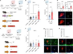
Recently at the site we saw this headline: "Baylor crowned STAT Madness champion for second consecutive year, for insights into memory." This is followed by the utterly untrue claim that "researchers showed brain cells called astrocytes are involved in forming and recalling memories." 

We read of something called the 2025 STAT Madness competition, "a bracket style celebration of neuroscience research." We read of some voting process, although we get no details of how this worked.  We read the claim that the competition "stacked 64 entries against each other in a month long combination."  Sadly the winner of this competition was a very low-quality piece of junk science entitled "Learning-associated astrocyte ensembles regulate memory recall." You can read the abstract here

The paper is behind a paywall. Why has the Statnews.com site given an award for a study that the public cannot even access? But without  paying any money, I can use the abstract link to get all the information I need to determine that the study is junk science.  For the abstract link does allow me to look at some of the paper's figures  And those figures tell me enough to determine the junk science nature of the paper. 

Figure 1 is below, and it has two "freezing percentage" bar graphs at its bottom.


Figure 4 and Figure 5 look similar. Figure 4 (entitled "Reactivation of LAAs elicits memory recall") has three "freezing percentage" bar graphs.  Figure 5 (entitled "Ensemble-specific NFIA is necessary for context-specific memory") has two "freezing percentage" bar graphs. 

From these figures, I can tell that the paper is guilty of two of the methodological sins so very common in memory-related rodent research, either one of which is enough to show the paper is very low-quality research.

Fault 1: Judgments of Freezing Behavior Were Used to Try to Judge Animal Recall

 We can tell from the figures mentioned above that the researchers attempted to measure recall in a rodent by using a "freezing behavior" estimation. All rodent experiments that use such a method are examples of junk science. The technique involves putting a rodent in a cage in which there is some stimulus the animal was trained to fear, and then trying to judge fear or recall by trying to judge what fraction of a time interval the rodent was immobile, with the immobility being judged as the rodent "frozen in fear." Neither fear nor memory nor recall can reliably be measured by such a technique. How often a rodent is immobile in a cage is a random thing that does not reliably correlate with how much the rodent is afraid or how often the rodent recalls a fearful stimulus.  For a full discussion of the utter unreliability of "freezing behavior" judgments in neuroscience experiments, see my long post here, entitled "All Papers Relying on Rodent 'Freezing Behavior' Estimations Are Junk Science." 

freezing behavior in rodents

I lived in a large apartment building for more than a decade, and about once a month I would spot a mouse, which would usually cause me to shriek. It seemed that not one time did I ever see a mouse freezing in fear after getting this fearful stimulus, contrary to the assumptions of neuroscientists assuming that mice tend to freeze in immobility when they are afraid. It would seem to always be the opposite behavior: the mice would always flee. Tracking the immobility of a mouse in a cage (and assuming the more "freezing" the more fear) is not a reliable way of measuring fear or recall in a mouse. 

Fault 2:Way-Too-Small Study Group Sizes

The use of way-too-small study group sizes is more the rule than the exception in today's neuroscience rodent research. 

The paper "Prevalence of Mixed-methods Sampling Designs in Social Science Research" has a Table 2 giving recommendations for minimum study group sizes for different types of research. According to the paper, the minimum number of subjects for an experimental study are 21 subjects per study group. The same table lists 61 subjects per study group as a minimum for a "correlational" study. The table below is a shortened version of the Table 2 found in that study. Because of the small effect sizes and "levels of significance" typically reported in neuroscience experiments, we should regard the sample sizes required for neuroscience experiments as every bit as high as the numbers listed below. 

Research design/method

Minimum sample size suggestion

Correlational

64 participants for one-tailed hypotheses; 82 participants for two-tailed hypotheses (Onwuegbuzie et al ., 2004)


Causal-comparative

51 participants per group for one-tailed hypotheses;

64 participants for two-tailed hypotheses (Onwuegbuzie et al., 2004)


Experimental

21 participants per group for one-tailed hypotheses

(Onwuegbuzie et al., 2004)


Phenomenological

<= 5/10 interviews (Creswell, 1998); >= 6 (Morse, 1994


For correlational, causal-comparative and experimental research designs, the recommended sample sizes represent those needed to detect a medium (using Cohen’s [1988] criteria), one-tailed statistically significant relationship or difference with 0.80 power at the 5% level of significance.”


Source: “Prevalence of Mixed-methods Sampling Designs in Social Science Research” by Kathleen M.T. Collins, Anthony J. Onwuegbuzie and Qun G. Jiao. 


In her post “Why Most Published Neuroscience Findings Are False,” Kelly Zalocusky PhD calculates that the median effect size of neuroscience studies is about .51. She then states the following, talking about statistical power (something that needs to be substantially greater than .5 for any compelling result to be claimed): 

"To get a power of 0.2, with an effect size of 0.51, the sample size needs to be 12 per group. This fits well with my intuition of sample sizes in (behavioral) neuroscience, and might actually be a little generous. To bump our power up to 0.5, we would need an n of 31 per group. A power of 0.8 would require 60 per group."

Here Zalocusky is telling us that the sample size requirements (i.e. study group size requirements) are just as high as those suggested in the table above, and that a study group size of around 50 is needed to get a good statistical power of 0.8 (80%). 

What study group sizes were used for the paper "Learning-associated astrocyte ensembles regulate memory recall," the paper that won the 2025 STAT Madness competition?  You can tell by the number of dots that we see at the bottom of the figures such as the Figure 1 at the top of this post (and also Figure 4  and Figure 5 of the paper). Each dot is a data point for a particular rodent. We see only about 8 dots for each of the study groups used. So the study group sizes were a way, way too-small size of only about 8 subjects per study group. 

freezing behavior bar chart

The study is therefore worthless as evidence, having used study group sizes less than half (and probably less than a third) of the study group sizes needed for a reliable result. StatNews.com (or the voters it coordinated) made the very bad mistake of giving a prize to a very low-quality junk science paper. 

How to Do Work Like This Junk Science Result

I can give an example of how you could do work similar to the utterly shoddy work that was awarded the STAT prize. You might do a project trying to show that some people can predict which cards are at the top of a newly shuffled deck that has been cut. 

A very important of your work would be to not publish beforehand any exact plan for how the research would be done.  Then you could do some tests that would work line this:

(1) You shuffle a deck of cards three times, and then cut the deck. 

(2) You bring in someone, asking him to guess the first ten cards that you will draw from the top of the deck.

You could have a "Correct Guesses" sheet recording the drawn result and the guess result for each of the guesses. Now, it would help very much if you used a technique that did not reliably record the guesses the guesser made. It might work like this: you could ask the user to list a sequence of ten guesses. You would then deal the first ten cards out on the table. You would then record your best recollection of the sequence of ten cards that the user had called out. 

This would not be a reliable measurement technique, because you had failed to write down each guess just after the guesser made it. So you would now be relying on your memory of the sequence of 10 cards the user chose. You could easily bias things by failing to remember correctly one or more of the cards the guesser had stated. This would help you get a more favorable "correct guesses" total. 

Now doing such a test on a few subjects, you would have some data: the sequence of ten cards drawn, and what the ten guesses made were (or at least, your best guess about what those guesses were, based on your short-term memory). You would then have a variety of ways to analyze your data. You could use the entire sequences of ten guesses.  Or if you did not like the "percentage correct" result from that, you could use only the first five cards guesses. Or if you did not like the "percentage correct" result from that, you could use only the last five cards guessed. Or if you did not like the "percentage correct" result from that, you could use only the middle five cards guessed. 

Or, if you were not getting anywhere trying to show above-chance card guessing, you could only report correct guesses of the card number or card face. Or, if you still were not getting anywhere trying to show correct card guessing, you could only report correct guesses of the card suit (clubs, spades, hearts or diamonds). Or, if you still were not getting anywhere trying to show correct card guessing, you could only report correct guesses of the card color (black or red). Or if the entire set of data showed no effect above chance for the card guessing, you could report on only an above-chance result for a single guesser. 

What happened if you tried all these things for 5 subjects, and were still unable to find anything greater than chance? No problem. You could simply "file drawer" the results, filing them away in your files. You could then start a new experiment using a different set of 5 subjects. After a few such attempts, you would probably have something you could report as an "above chance" result for card guessing, given your small study group sizes, and given your freedom to analyze the data in any of dozens of different ways. False alarms tend to show up with small data sets. Most of those false alarms go away when you use a much larger study group size. 

For example, if you flip a coin 100 times you will get a number of heads very close to 50. But if you flip a coin only a small number of times, it is very easy to get a result much different from the result expected by chance. So, for example, it is not too hard to flip a coin six times and get four or more heads or four or more tails rather than the chance-expected result of three heads. The chance of such a result is greater than 33%. Do three trials of six flips each, and you will probably get one trial with four heads or four tails. 

The example given above is very much like the typical neuroscience rodent experiment. Just like the card-guessing experiment I described, a typical neuroscience experiment fails to publish in advance any detailed research plan, leaving the experimenters free to analyze data in any of innumerable ways. For example, there are no standards for how to judge "freezing behavior" in rodents. So an experimenter can attempt to judge how much a rodent was immobile after being exposed to some fear stimulus it was trained to fear, recording three minutes of data. If the experimenter does not like the "freezing percentage" recorded for the first three minutes, the experimenter can simply report on only the "freezing percentage" recorded for the first two minutes. If the experimenter does not like the "freezing percentage" recorded for the first two minutes,  the experimenter can simply report on only the "freezing percentage" recorded for the first 60 seconds. If the experimenter does not like the "freezing percentage" recorded for the first 60 seconds, the experimenter can simply report on only the "freezing percentage" recorded for the first 30 seconds. Since there is no convention of a standard time interval to use, and since researchers typically get away without even reporting what was the time interval corresponding to a "freezing percentage" graph, it is easy for a researcher to switch around the time interval in any way he wants, and not even use the same time interval for each freezing percentage" graph in his paper.

And just as the card-guessing experimenter I described used an unreliable technique for measuring a key element of the experiment (relying on the experimenter's memory of what 10 called guesses were), nowadays neuroscience rodent memory experiments rely on an utterly unreliable measurement technique: the technique of trying to judge an animal's recall by judging an animal's immobility during an arbitrary span of time, with that immobility called "freezing behavior." There are reliable ways of measuring whether a rodent recalled something. You can use heart-rate measurement to measure heart rate spikes, and that is a reliable way of telling whether a rodent is recalling a fearful stimulus (the heart rate of rodents spikes dramatically when they are afraid). Or you can use something like the Morris water maze, widely regarded as a reliable way of measuring how well a rat remembered something. Or you can use the Fear Stimulus Avoidance technique described below. 

reliable neuroscience recall measurement

But if you use the unreliable "freezing behavior" technique, your paper deserves nothing but scorn. And the more "freezing behavior" graphs that your paper has, the more it deserves scorn and contempt. 

Nowadays neuroscience memory research involving rodents tells us mainly one important thing about memory: that neuroscience rodent researchers seem to have memories so bad that they keep forgetting to follow good standards in doing research. 

bungling neuroscientist

bungling neuroscientist

bungling neuroscientist

bungling neuroscientist

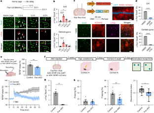
Postscript: The same fatal defects of the paper discussed above are found in the recent junk science paper "EPSILON: a method for pulse-chase labeling to probe synaptic AMPAR exocytosis during memory formation." The paper uses way too-small study group sizes of only 3 mice, 4 mice and 6 mice. The paper also hinges upon a use of the totally unreliable method of trying to judge "freezing behavior" to try and judge whether fear recall occurred in mice. The longer the time interval over which so-called freezing behavior is judged, the less reliable is the claim to have measured fear or recall, because real "freezing behavior" would be a very short-lasting phenomenon, probably lasting less than 30 seconds. In this case the "freezing behavior" is judged over a length of three minutes, a particularly dubious length of time to be using when such a judgment about "freezing behavior" is done. 

We again have an example of the arbitrary decisions and lack of standards that go on when such "freezing behavior" judgments are made, the kind of deal in which any experimenter is free to use any criteria, in a "see whatever you want to see" manner. The authors tell us that any motion of less than .03 meters per second (about less than one inch per second) was counted as "freezing behavior," stating, "the time duration during which the speed was slower than 0.03 m s−1 was counted as freezing time."  That makes no sense. Moving .8 inch a second is not actually freezing behavior for a mouse. Freezing behavior is immobility.  

The authors failed to do a sample size calculation to determine the number of mice needed to get a result of decent statistical power. Rather than confess their failure to do this basic requirement of good experimental science (a sample size calculation), the authors strangely state, "Sample sizes were determined by the technical requirements of the experiments." That sounds like someone saying the equivalent of, "We couldn't use more mice because it would have been too much trouble," which is very lame. The authors do candidly confess, "Data collection was not conducted blinded to experimental conditions." No paper of this type should be taken seriously unless the authors followed a detailed blinding protocol, and the authors did no such thing. 

Proving that you can't trust anything merely because it appears in a Harvard publication, the Harvard Gazette has published a recent article boasting about this piece of very bad junk science, one with the bogus headline, "Tracking precisely how learning, memories are formed." Nothing of the sort was done, because of the very bad methods followed.