If you do a Google search using the phrase "memory research fraud," you will now get many recent results from leading news sources, including results such as these:
- What allegations of Alzheimer's research fraud mean for patients
- Whistleblower Lifts the Lid on “False” Alzheimer's Research
- Research Fraud Bombshell Threatens Amyloid Theory of Alzheimer's Disease
- Decades Of Alzheimer’s Research Could Be Based On Fraudulent Data
- DECADES OF ALZHEIMER'S RESEARCH ALLEGEDLY BASED ON FABRICATED DATA
The leading journal Science did a big six-month investigation resulting in a recent long article entitled "Blots on a Field?" We hear claims of doctored images and fake visuals. An interesting part of the story is where the journal Science reaches out to leading science journals that have allegedly published some fake research, such as Nature and the Journal of Neuroscience, getting a lot of "no comments" from such journals.
We read this in one news article:
"Dr Bik has now identified 14 other studies...that also appear suspicious. Despite this, in the majority of cases, no action has been taken against the journals that published them. The University of Minnesota declined a request to comment by The Mail on Sunday...Richard Smith, a former editor-in-chief of the British Medical Journal (BMJ), who has warned that research fraud is a ‘major threat to public health’, said that the case was ‘shocking but not surprising’. He cites research that suggests up to one in five of the estimated two million medical studies published each year could contain invented or plagiarised results, details of patients who never existed and trials that did not actually take place. He adds the problem is ‘well known about’ in science circles, yet there is a reluctance within the establishment to accept the scale of the problem."
The same Dr. Bik is quoted as saying, "’I've flagged more than 6,000 studies as potentially fraudulent, but just one in six have been retracted by publishers." Later in the same article we read this:
"At present there are no drugs that can fight Alzheimer’s. The first company to invent one would no doubt have a billion-dollar blockbuster on its hands – and this, says Adrian Heilbut, has incentivised misconduct."
We can imagine part of the motivation here:
(1) Invest money in company XYZ.
(2) Do a "fair means or foul" paper suggesting that company XYZ's approach towards treating Alzheimer's is promising.
(3) Watch your stock shares soar in value.
The items discussed above are only "the tip of the iceberg." The problem in memory research is ten times worse than the mere problem of some researchers doing image doctoring to produce frauduent images. The problem that is ten times worse involves things like this:
(1) Scientific papers are routinely stating claims in their titles and abstracts that are not well-established by any observations reported in the papers.
(2) Such unfounded claims are being massively repeated in the uncritical "echo chamber" that is the mainstream press and body of web sites calling themselves "science news" sites.
(3) Scientists doing experiments involving memory typically use study group sizes that are too small to produce any reliable result. The results are mainly false alarms of a type that can easily arise when too-small study group sizes are used.
(4) Scientists doing experiments involving memory typically fail to do the sample size calculations that would alert them that the study group sizes they are using are way too small to produce a reliable result.
(5) Scientists doing experiments involving memory are very often using defective experimental procedures that produce unreliable results, such as trying to measure fear in rodents by subjectively judging "freezing behavior" rather than using better procedures producing more reliable results, such as trying to measure fear in rodents by measuring heart rate (which reliably spikes very sharply when a rodent is afraid).
(6) Scientists doing experiments involving memory routinely fail to follow a blinding protocol that would reduce the chance of them producing false-alarm results in which they merely "see what they want to see."
(7) Scientists doing experiments involving memory routinely fail to follow good practices by pre-registering an exact experimental method for collecting and analyzing data. Often their papers show strong signs of "keep torturing the data until it confesses," which can also be described as "keep slicing and dicing the data until you find something like you hoped to find."
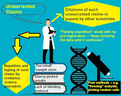
The diagram below illustrates some of what is going on. The "picking random cells" refers to memory experiments in which some learning occurs, and then scientists attempt to show neural changes resulting from learning after randomly picking some cells for analysis, ignoring the extreme improbability that randomly selected cells would have changed because of such learning. Because of constant remodeling and molecular turnover occurring theoughout the brain, randomly selected cells or synapses will be likely to show changes that were not produced by learning.
The links at the top of this blog refer to a scandal involving misleading images in neuroscience papers. Something similar has gone on endless times in brain imaging studies on the neural correlates of consciousness. Again and again, such studies will show visuals that depict differences of only 1% or smaller between brain activity in different small regions of the brain. But such regions will be shown as red regions in brain images, with all of the other areas having a grayish “black and white” color. When you see such an image, you inevitably get the impression that the highlighted part of the brain has much higher activity than other regions. But such a conclusion is not what the data is showing.
So, for example, a study finding merely 1% higher brain activity in a region near the corpus callosum (under some activity that we may call Activity X) might release a very misleading image looking like the image below, in which the area of 1% greater activity is colored in red.
But such an image is lying with colors. If there is only a 1% greater activity in this region, an honest diagram would look like the one below.
With this diagram, the same region shown in red in the first diagram is shown as only 1% darker. You can't actually tell by looking at the diagram which region has the 1% greater activity when Activity X occurs. But that's no problem. The diagram above leaves the reader with the correct story: none of the brain regions differ in activity by more than 1% when Activity X occurs. Contrast this with the first image, which creates the very misleading idea that one part of the brain is much more active than the others when Activity X occurs.
You might complain that with such a visual, you cannot tell which regions have the slightly greater activity. But there are various ways to highlight particular regions of a brain visual, such as circling, pointing arrows, outlining, and so forth. For example, the following shows a region of very slightly higher activity without misleading the viewer by creating the impression of much higher activity:
The misleading diagrams of brain imaging studies seem all the more appalling when you consider that the images in such studies are typically the only thing that laymen use to form an opinion about localization in the brain. The text of brain imaging studies is typically written in thick jargon that only a neuroscientist can understand. Frustrated by this very hard-to-understand jargon and unclear writing, every layman reading these studies forms his opinions based on the visuals. When such visuals deceive us by lying with colors (as they so often do), it is a scandal of visual misrepresentation as great as whatever is discussed in the links at the top of this post.


No comments:
Post a Comment