Tuesday, July 21, 2020

Preservation of Mind and Memories After Removal of Half a Brain

"Scientific journals have reported cases of persons whose injuries have necessitated the removal of a large portion of the brain, and whose memory and power of thought were unimpaired by the loss of much cerebral matter, or by damage to centers which are supposed to be necessary to memory and consciousness. Dr. Troude writes:  'As M. Bergson foresaw in 1897, the hypothesis of the brain as conservative of memory images, must be renounced once and for all, and other ideas as to the nature of its role in the act of memory must be accepted. ' "

--- Helen C. Lambert, A General Survey of Psychical Phenomena, p. 49.

The idea of a crucial experiment or critical experiment is an old concept in the world of science. Such an experiment is supposedly one that leaves one particular hypothesis standing, and rules out all rival explanations or rival hypotheses. The idea that there are such experiments has been criticized by some. A simpler idea is the idea of a sink-or-swim experiment. A sink-or-swim experiment is one that either leaves some hypothesis standing as a viable hypothesis (the “swim” situation) or causes the hypothesis to be discredited (the “sink” hypothesis).

Scientists have very often claimed that the human mind is produced by the brain, and that memories are stored in the brain. A very interesting question is: could you do a sink-or-swim experiment testing such hypotheses? The experiment has actually been done, not just once but many times. I will here use the term “experiment” for medical procedures that were usually done for medical reasons such as stopping very bad brain seizures in patients. Although the doctors who did such procedures may not have considered them experiments, we can consider them as experiments in the sense of testing a particular hypothesis about the brain.

The sink-or-swim experiment for the hypothesis that the brain makes the mind and the hypothesis that the brain stores memories is to surgically remove half of the brain, and see what the effect is on the mind and memory. Such an experiment has been done many times. Almost every time the result has been that there was no major effect on consciousness, no major effect or intelligence, and no major effect on memory. The memories of people who had half of their brains removed usually preserved the knowledge and life memories they had acquired.

This is a “sink” result for this sink-or-swim experiment. The results of such surgical operations decisively refute claims that the mind is the product of the brain and claims that the brain is the storage place of memories. But addicted to materialist dogma that the mind is merely the product of the brain and that memories are stored in brains, virtually no neuroscientists have paid attention to the results of these sink-or-swim experiments. In this regard, they are like fundamentalists who keep believing that the Earth is 6000 years old despite observational results indicating our planet is billions of years old.

I have in five previous posts (here, here, here, here and here) listed very much data relating to such experiments. In this post I will not restate that data showing that intelligence is well-preserved after removing half of the brain, but will mostly cite some data and cases I have not previously discussed.

I can start with the results reported in the American Journal of Psychology, Vol. 46, No. 3 (Jul., 1934), pages 500-503, regarding work of W. E. Dandy, in which he removed half of the brains of patients. You can read the results in the preview here (without doing any registration). We read the following (I have put a few of the sentences in boldface):

Dandy has completely removed the right cerebral hemisphere from eight patients. He has performed total extirpations of one or more lobes much oftener... There are tabulated below certain generalizations on the effects of removing the right hemisphere.... The operation was the complete extirpation of the right frontal, temporal, parietal, and occipital lobes peripheral to the corpus striatum. The weight of the tissue re moved varies, with the pathological conditions involved, from 250 to 584 grm [grams].Coherent conversation began within twenty-four hours after operation, and in one case on the afternoon of the same day. Later examinations showed no observable mental changes. The patients were perfectly oriented in respect of time, place, and person; their memory was unimpaired for immediate and remote events; conversation was always coherent; ability to read, write, compute, and learn new material was unaltered. Current events were followed with normal interest. There were no personality changes apparent; the patients were emotionally stable, without fears, delusions, hallucinations, expansive ideas or obsessions, and with a good sense of humor; they joked frequently. They showed a natural interest in their condition and future. They cooperated intelligently at all times throughout post-operative care and subsequent testing of function.”

It would be rather hard to imagine a more decisive refutation of the claim that the human brain is the source of the human mind, and the claim that the human brain is the storage place of human memories. Here are eight people who had half of their brains removed. Yet the people showed “no observable mental changes,” and “their memory was unimpaired for immediate and remote events.” The people could read, write, compute and learn just as if nothing had happened, and “there were no personality changes.”

A 1966 paper was entitled “Long-term changes in intellect and behavior after hemispherectomy.” The paper refers to operations in which half of a brain is removed, often to stop very bad brain seizures. This paper gives very detailed “before and after” IQ score data on 11 people who had half of their brains removed. Eight of the 11 people had the left half of their brain removed, and the other three had the right half of their brain removed. Every single one of the 11 people was able to get an improved IQ score on at least one of the tests taken after half of their brain was removed, a score better than a corresponding score they had got before half of their brain was removed.

Patient 1 (a P.G.) had an IQ of 128 before half of his brain was removed. After half of his brain was removed, he scored 142 on an IQ test. The paper tells us that this man with half a brain “obtained a university diploma after operation” and “has a responsible administrative position with a local authority.”

The same paper refers to previous results when removing half of a brain, and notes data suggesting that such an operation has little negative effect on intelligence. Referring to intelligence, we are told that McKissock reported “short term improvement in 13 of 17 cases,” that another researcher found “significant improvement in verbal intelligence scores in a variety of tests after operation in five of 35 cases, with temporary deterioration in two, the remainder unchanged.” We are also told that White “reports improvement in personality in 80% of 134 cases” in which half of the brain was removed.

The 2013 paper "Long-term functional outcomes and their predictors after hemispherectomy in 115 children" reports this: "In this cohort of 115 children, at a mean follow-up of 6.05 years after hemispherectomy, 83% patients walked independently, 73% had minimal or no behavioral problems, 69.5% had satisfactory spoken language skills, and 42% had good reading skills."  These results are for people with half-brains, and we should remember that a large fraction of people with full brains lack good reading skills.  Typically a hemispherectomy operation occurs as a last resort for a child who has been long-plagued by seizures.  Before the operation, such seizures may have long-disrupted normal learning.  So this 42% may not even reflect any major deterioration in reading skills after removal of half a brain. 

In the scientific paper here, we have on page 248 and page 250 before and after test scores for various subjects who had of their brains removed in hemispherectomy operations.  The IQ score differences are slight. IQ tests don't involve learned information, but almost any IQ test would be largely a test of memory, as it would be a largely a test of ability to read test questions.  

On the same pages we have before and after test scores for Peabody Picture Vocabulary Tests given to various subjects who had  half of their brains removed in hemispherectomy operations.   In these tests, someone is shown picture cards like the one below, and asked to name the words represented by the pictures.  These tests are tests of memory retention after removal of half of the brain.  On these memory tests there was no decline in the score of 21 subjects mentioned on page 248, and no decline in 7 subjects mentioned on page 250. 


In an article in the New Yorker magazine, we are told of a Christina Santhouse who had half of her brain surgically removed: “When I met her, she had taken her S.A.T.s and just finished high school, coming in seventy-sixth in a class of two hundred and twenty-five.” If your brain makes your mind, how could you finish in the top 34% of your class with only half a brain? The same article tells us of someone who had half of the brain removed, but made the dean's list in college, a list of the top-performing students on campus.

An article in the LA Times tells us about memory preservation in a young girl who lost half her brain:

How is it that 8-year-old Beth Usher of Storrs, Conn., can lose her left hemisphere, yet retain her large repertoire of knock-knock jokes? Beth’s memories survived not just the loss of brain tissue, but also the 32 days that she spent in a coma, the result of some brain stem swelling that occurred in response to the trauma of surgery. Shortly after Beth regained consciousness, her father began quizzing her about people and places from her past. Brian Usher didn’t get very far. 'Dad,' Beth interrupted, with a trace of impatience. 'I remember everything.' ”

In a scientific paper ("Why Would You Remove Half a Brain? The Outcome of 58 Children After Hemispherectomy −−The Johns Hopkins Experience: 1968 to 1996") we read about how surgeons at Johns Hopkins Medical School performed fifty-eight hemispherectomy operations on children over a thirty-year period. Eleven of these children had the left hemisphere of their brains removed; most of the rest had the right hemisphere of their brains removed.  The paper states this:

"Despite removal of one hemisphere  [i.e. one half of the brain], the intellect of all but one of the children seems either unchanged or improved....Although there have been major concerns about loss of language after left hemispherectomy, all eleven of these children have regained virtually normal language....It is tempting to speculate, that the continuous electrical activity of these severely dysfunctional hemispheres interferes with the function of the other, more normal hemisphere. This might explain why motor function improves after hemispherectomy and why language recovers after removal of the dysfunctional left hemisphere, but does not seem to fully transfer before surgery. Perhaps it also partially explains intellectual improvement in these children after removal of half of the cortex. We are awed by the apparent retention of memory after removal of half of the brain, either half, and by the retention of the child’s personality and sense of humor."

There is a reason why we can be confident that removal of half of a brain in hemispherectomy operations does not cause any major loss of learned memories.  If there was a case of any such thing happening, you can believe that it would be endlessly recited by those who wish for us to believe that memories are stored in brains.  But there is no such case, so we never hear materialists telling us about some person who suffered some dramatic loss of learned knowledge after having a hemispherectomy operation in which half of his brain was removed. 

Our professors very often make biology claims that are contrary to the low-level facts of biology. The table below lists various cases in which the fantasy biology of academia dogma diverges from biology reality. 

FANTASY BIOLOGY VERSUS BIOLOGICAL REALITY
Dubious Biology Claim Biological Reality
Brains store memories, probably in synapses or dendritic spines. Neither synapses nor dendritic spines last for even a tenth of the longest time that humans can remember things, and both are made up of proteins with lifetimes of only a few weeks.
DNA stores a blueprint or recipe for making the human body. DNA does not specify the physical structure of any of these things: an organism's body, its organ systems, its organs or its cells. 
Visible biological innovations arise from a combination of random mutations and natural selection, which improves the DNA of a species. It has not been proven that any visible complex biological innovation ever appeared because of random mutations and natural selection, and we know of a reason why mere DNA mutations could never produce a complex visible biological innovation: that visible physical structures are not specified in DNA.
Life appeared because of a lucky combination of random chemicals billions of years ago. Neither a living thing nor any of the building blocks of a living thing (proteins and nucleic acids with genetic information) has ever been produced through any experimental process that  realistically simulated early Earth conditions.
The building blocks of life have been found in outer space. No one has found in outer space either of the two actual building blocks of life: proteins or nucleic acids with genetic information.
Brain scans show your brain makes your mind. Brains scans actually show signal differences of less than 1% during thinking or recall, what we would expect from random variations.
Brain signals are real fast. Synaptic delays, synaptic fatigue and relatively slow dendritic transmission mean that signals in the cortex must be real slow.
The common descent of all life from a single ancestor is a fact. A shortage of transitional fossils and the lack of DNA corresponding to old fossils (because of DNA's half-life of 521 years) make the doctrine of common descent very unproven.
Chemically humans are almost exactly like chimps. 80% of proteins are different between humans and chimps.
Our minds can be explained neurally. There is no credible neural explanation for any of the main features of the human mind: memory, self-hood, consciousness, abstract thinking, and imagination.
We kind of understand how a speck-sized egg can progress to become a full-sized baby. We have no understanding of how this occurs (given a lack of a body plan in DNA), and do not even understand what causes cells to reproduce.
Memory and intelligence depend strongly on brain status. A person can lose half of his brain in a hemispherectomy operation, with little effect on memory or intelligence.

The image below reproduces the table above. 


biology myths

Postscript
: The paper "
Language recovery after hemispherectomy in children with late-onset seizures" gives us before-and-after IQ scores for six children having removal of the left half of the brain (Table 1, FSIQ row). Such comparisons can be affected by short-term deficets that later are reduced. The comparison shows one child dropped 20 IQ points, one child dropped 10 IQ points, another child gained 3 IQ points, another child dropped 15 IQ points, another child gained 11 IQ points, and another child had no change in IQ. The very mixed results are consistent with the brain not being the source of the human mind. 

half a brain but good mind

On page 254 of the textbook here, we read a statement by someone saying that he performed 43 hemispherectomy operations. He says, "The secondary outcome scores reevaluated at six months and 1 year showed no significant difference in IQ," indicating intelligence is not affected by removal of half of the brain. 

Page 5 of the scientific paper here describes a 7-year-old girl who is "fully bilingual in Turkish and Danish" despite having had most of half of the left side of her brain removed in a hemispherectomy operation at the age of 3. We are told that except for a slight spasticity, "she leads an otherwise normal life." 

On page 3 of the Florida newspaper you can read here, the Lake City Reporter of December 25, 2015,  we have a quote by brain surgeon Ben Carson:

"He recalled meeting one patient to whom he’d
given a hemispherectomy — removing half the brain
— as an infant. 'He had graduated from college
number one in his class — with half a brain,' Carson
said." 

Below is an excerpt from the 1931 newspaper article "How Much Brain Used?" by William Brady M.D.:

"Aside from the fact that the boy felt no pain from the surgical treatment (and had no anesthetic), it was remarkable that he retained full consciousness and unimpaired mental function, al| though a considerable part of the cerebrum was destroyed. Recent surgical observation has proved that the whole of the right cerebral hemisphere may be removed without disturbance of mentality: this has been done repeatedly for the removal of large brain tumor. Both right and left frontal lobes of the brain may be removed without disturbing the patient's mentality. The patient remains perfectly oriented as to time, place and person; the memory is unimpaired: reading. writing and mathematical calculation are still done accurately; conversation is normal. In other cases the left occipital lobe and the lower third of the left temporal lobe have been removed without apparent. effect on mentality. "

You can read the full article using the link below:

https://chroniclingamerica.loc.gov/lccn/sn83045462/1931-01-20/ed-1/seq-28/

Below is the first page of a 1933 Popular Science article written by a doctor, one you can read here. We are told of a woman who had the "entire right half" of her brain removed. We are told that a few hours after the operation she "recognized and talked with her friends." The operation apparently caused no loss of learned knowledge or memories.  Both the act of recognizing a friend and the act of talking requires the use of learned knowledge such as the knowledge of what a friend looks like, and the knowledge of the words in a language and the grammar rules of the language. We are told the woman "improved rapidly, reading and writing during her convalescence." No mention is made of any loss of memory.  The reported result is consistent with the idea that the brain does not store memories. 

preservation of memory after hemispherectomy

The next page mentions several case histories of people who suffered the most horrible brain injuries in various types of accidents. No mention is made of any damage to memory or mind. We hear instead comments such as "apparently his mind did not suffer from the accident" and mention of a "miraculous recovery."

Another case is discussed here, in a book chapter of a textbook. We read of a patient E.C. who had a large brain tumor. To treat the tumor, the left half of the patient's brain was removed, in a hemispherectomy operation. We read that "the entire hemisphere was removed in one piece" on December 7, 1965. We read that "immediately following hemispherectomy" the patient had an "ability to follow simple verbal commands."  We read on page 107 that "the patient spontaneously articulated words and short phrases fairly well immediately after the operation."  

We read that this patient E.C. "gave the correct number of years when asked how long he had lived at his house." We read on page 107 that on January 26, 1966 (about six weeks after the operation) he "slowly but correctly selected four of the first six items from six pages, each with four items to choose from, before indicating he was tired" when given the Peabody picture vocabulary test. Such a test involves a tester speaking a word, and the user pointing to the correct picture in a grid of 4 pictures. 

The image below (from the paper here) shows an example of one of the pages. The word spoken by the tester is "citrus."

The result described above (4 out of 6 correct on six such pages) clearly suggests that patient E.C. retained his learned knowledge after the left half of the brain was removed. Since the test was given only six weeks after the operation, and E.C. was in a recovering state during those weeks in which he was not being schooled, we cannot explain such a result as being due to knowledge acquired during those six weeks. We also read on page 107 that on June 3, 1966 patient E.C. correctly named 85 out of 112 items on such a 
Peabody picture vocabulary test. No mention is made of schooling occurring during the first six months of 1966. 

On page 109 we read of three other cases of the removal of the left half of the brain, and we read that "speech and verbal comprehension were present immediately after left hemispherectomy in all three cases." We read nothing about any loss of knowledge or episodic memories in any of these cases.  Neither speech nor verbal comprehension can occur unless a great deal of learned knowledge is retained. 

The news story here documents a very clear case of the preservation of memory after the removal of the left half of the cerebrum. We read of a 47-year-old man who had the left half of his cerebrum removed. We read this:

"Dr. Smith said the patient's memory of events before the operation 'was well preserved, much better than anyone expected.' Tests following surgy show Coe can perform many functions traditionally thought impossible after removal of the brain's dominant hemisphere. Immediately following surgery, Coe still had the ability to speak spontaneously."

A report like this does not merely contradict the claim that the brain is the source of the mind. It also contradicts the claim that the brain is a storage place of memories. 

The 1963 newspaper account here is entitled "Monkeys With Half a Brain." We read this:

"Fifty monkeys have had half of their brains removed. Their memories appear to be as complete as before surgery. Until this experiment was made, even doctors thought that such surgery would affect memory....Not only is memory retained; the monkeys are alert, intelligent, sociable, and have the same enthusiasm as doing things as before....What are some of things the team has discovered from observation of monkeys with half their brain removed? The monkeys remember how to do things taught them before surgery. Such as which of two blocks of wood place under them has a reward under it -- a raisin or other food. Fears are recalled....'While the rhesus monkeys we test remember everything taught them before surgery, it takes them twice as long to learn something new as monkeys with both half of their brain,' explains Dr. Kruper. 'But the important thing is that they DO learn, even if it does take them longer.' "

The visual below "speaks volumes" on this topic. It is the result of a search on Google Scholar for "amnesia after hemispherectomy." "Hemispherectomy" is the standard name for the operation that has been performed thousands of times, in which half of the brain is removed to stop very bad seizures. "Amnesia" is the main term used for a loss of episodic memories.  Any search on Google Scholar will produce very many results when there are quite a few cases corresponding to such a search. For example, doing a Google Scholar search for "vision loss after automobile accident" produces many results. But doing a search for "amnesia after hemispherectomy" produces zero results.

amnesia after hemispherectomy

The results discussed in this post deserve to be pondered at great length. The results indicate very strongly that the human brain is not any storage place of human memories. After considering such results, you should also ponder the most important null result of neuroscience research: the fact that brain tissue from thousands of living and recently deceased humans has been microscopically studied at great length, with the most powerful microscopes; but no one has ever found the slightest trace of learned information or any episodic memory by such microscopic examination. No one found a single phrase, a single word or even a single letter of the alphabet after microscopically examining brain tissue. Nor did anyone ever find by microscopic examination of brain tissue any trace of anything a human ever saw or heard. 

Thursday, July 9, 2020

Gender Differences in Brains Help Discredit Prevailing Dogmas About Brains

Many people are interested in differences between the brains of males and the brains of females, and differences between males and females in IQ tests and memory tests. A careful examination of this area provides some evidence against the claim that the brain is the source of human intelligence, and the claim that memories are stored in synapses of the brain.

The brains of males are significantly larger on average than females -- about 10% bigger. But we know that females tend to be shorter and weigh less than males. Some say that the relative size of female brains (female brain sizes compared to female body sizes) is no smaller than the relative size of male brains.  But in a scientific paper a scientist states, "After correcting for body height or body surface area, men's brains are about 100 g heavier than female brains in both racial groups."  That difference of 100 grams is about 7% of the total weight of a male brain (about 1350 grams). 

So using the idea that the human mind is produced by the brain, we should expect that males do about 7% better at school and about 7% better in IQ tests.  But this is not at all the case. Males and females do about the same on IQ tests, with a difference of less than 1% or 2%.  In the United States females tend to get just as high academic grades as males.  In this regard, the claim that the brain is produced by the mind fails the observational test. 

Now let's consider human memory. The standard academic dogma (unsupported by any facts) is that memories are stored in the synapses of brains. The persistence of this dogma is mystifying, given what we know about the instability of synapses. Humans can reliably remember things for longer than 50 years, but individual synapses do not last for years. The proteins that make up synapses are very short-lived, having an average lifetime of only a few weeks. 

Wikipedia.org states, "Multiple studies[22] [23] have found a higher synaptic density in males: a 2008 study reported that men had a significantly higher average synaptic density of 12.9 × 10⁸ per cubic millimeter, whereas in women it was 8.6 × 10⁸ per cubic millimeter, a 33% difference." The 2008 study mentioned is the study "
Gender differences in human cortical synaptic density" you can read here

Now, this 33% difference is quite a big difference, much bigger than the brain size difference previously mentioned. Under the assumption that synapses are the storage place of memory, we should expect (given this 33% greater synapse density in males) that either males tend to have stored much more memories than females, or that males are better at remembering things than females. But  such things are not true. 

There is no evidence that males store more memories than females. One good way of testing whether males store more memories than females is simply to look at academic scores. If males tended to store more memories, they would tend to have higher academic scores than females. But females do just as well as males in tests of learned information. 

Below is a quote from an article in the New York Times indicating that boys do not do better than females (on average) in school tests:

"The study included test scores from the 2008 to 2014 school years for 10,000 of the roughly 12,000 school districts in the United States. In no district do boys, on average, do as well or better than girls in English and language arts. In the average district, girls perform about three-quarters of a grade level ahead of boys. But in math, there is nearly no gender gap, on average. Girls perform slightly better than boys in about a quarter of districts...Boys do slightly better in the rest."

Here are some quotes from the scientific paper "The Role of Sex in Memory Function: Considerations and Recommendations in the Context of Exercise": 

"Females tend to outperform males in episodic memory function....Females tend to perform better than males in verbal-based episodic memory tasks, as opposed to spatial-based memory tasks []. Females generally access their memories faster than males [], date them more precisely [], and use more emotional terms when describing memories []. Superior verbal memory for females also appears to be independent of intelligence level []. Additionally, females also have greater specificity for events imagined to occur in the future []. In general, females outperform males on autobiographical memory (particularly with high retrieval support via verbal probing []), random word recall [], story recall [], auditory episodic memory [], semantic memory (driven by superiority in fluency) [], and face recognition tasks [,]."


So the paper is telling us that female memory performance is better than male memory performance in all these areas. But how can that be, if males have a synaptic density 33% greater? We have here additional evidence that there is no truth in the common claim that memories are stored in human synapses. 

Sunday, June 28, 2020

Long Article Tries to Show Neural Memory Storage, but Gives No Real Evidence for It

In Discover magazine, there was recently a long article entitled “What Happens in Your Brain When You Make Memories?” An article like this is an attempt to convince us that scientists have some good understanding of how a brain could store memories. But the article completely fails at such a task, and provides no substantial evidence for any such thing as neural memory storage.

We are told the following: “In the 1990s, scientists analyzed high-resolution brain scans and found that these fleeting memories depend on neurons firing in the prefrontal cortex, the front part of the brain responsible for higher-level thinking.” There is no actual evidence that the front part of the brain is responsible for high-level thinking. You can read here for evidence that specifically contradicts such a claim. 

The quote above includes a link to a brain scanning scientific paper. That paper provides no evidence that memories depend on neurons firing anywhere. In any type of brain scanning study, the two main questions to ask are:  how many subjects were used, and what was the percent signal change detected during some supposed activation of some brain region? The paper does not tell us either of these things. It mentions some brain scanning study, but does not tell any details of how many subjects it used, or what percent signal change was detected. We can only assume that the study was one of those ridiculously common studies that either: (1) used too small a sample size to get a result of good statistical power, or (2) detected only meaningless signal changes such as less than 1%, the type of differences we would expect to get by chance, or (3) had both of these problems. When scientists use impressive sample sizes or when they get impressive brain scanning results regarding percent signal changes, they almost always tell us about such a thing. When there is a failure for a paper to mention either of these numbers, we should assume it is because the numbers were not impressive, and not good evidence.

The article then states the dogma that memories form when synapses are strengthened: “When a long-term memory is formed, the connections between neurons, known as synapses, are strengthened.” There is no evidence that this is true. When stating the sentence above, the article has a link to a paper that provides no evidence that memory storage involves synapse strengthening.

In fact, there are reasons why it cannot be true that memories are formed by synapses being strengthened. The first is that synapses are too unstable to be a permanent storage place for memories. The proteins in synapses have an average lifetime of only a few weeks. But humans can accurately remember things for 60 years, which is 1000 times longer than 50 weeks. Synapses do not last for very long. The paper here says that half-life of synapses is "from days to months." The 2018 study here precisely measured the lifetimes of more than 3000 brain proteins from all over the brain, and found not a single one with a lifetime of more than 75 days (figure 2 shows the average protein lifetime was only 11 days). 

The second reason is that humans are able to instantly form permanent new memories at a rapid clip. This was shown in an experiment in which humans were able to remember fairly well images they were only exposed to for a few seconds. The experiment is described in the scientific paper “Visual long-term memory has a massive storage capacity for object details.” The experimenters showed some subjects 2500 images over the course of five and a half hours, and the subjects viewed each image for only three seconds. Then the subjects were tested in the following way described by the paper:

"Afterward, they were shown pairs of images and indicated which of the two they had seen. The previously viewed item could be paired with either an object from a novel category, an object of the same basic-level category, or the same object in a different state or pose. Performance in each of these conditions was remarkably high  (92%, 88%, and 87%, respectively), suggesting that participants successfully maintained detailed representations of thousands of images."

Let us imagine that memories were being stored in the brain by a process of synapse strengthening. Each time a memory was stored, it would involve the synthesis of new proteins (requiring minutes), and also the additional time (presumably requiring additional minutes) for an encoding effect in which knowledge or experienced was translated into neural states. If the brain stored memories in such a way, it could not possibly keep up with remembering most of thousands of images that appeared for only three seconds each.

In the Discover magazine article, we are then told an inaccurate legend of scientific achievement: “ In a 2012 Nature study, Tonegawa and researchers at MIT and Stanford University used optogenetics to demonstrate that our memory traces do indeed live in specific clusters of brain cells.” No, Susumu Tonegawa and his colleagues did not do any such thing. In the post here you can read a rather lengthy discussion of various memory-related papers authored by people working at Tonegawa's MIT memory laboratory. These papers suffer from a common defect of using too-small sample sizes. Again and again when looking up the memory-related papers authored by people working at Tonegawa's MIT memory laboratory, I found papers that used sample sizes so small that were not good evidence for anything. In a neuroscience experiment, the absolute minimum for a somewhat compelling result is 15 animals per study group (and in most cases the number of animals per study group should be much higher, such as 25 or more). But again and again when looking up the memory-related papers authored by people working at Tonegawa's MIT memory laboratory, I found papers that used sample sizes of 10 or smaller. Such papers are not good evidence for anything.

In the Discover magazine article, we have a clear description of the utterly fallacious experimental technique used by Tonegawa, a technique that has given him the wrong idea that he has found a memory in brains. Here is what the article says:

"In the paper, the research team describes how they pinpointed a particular group of neurons in the hippocampus, a part of the brain involved in the formation of long-term memories, that start firing under certain conditions. In this case, the researchers did so by having mice explore an unfamiliar cage. '[Then] you give [the mouse] mild electric shocks to their footpads,' says Tonegawa. 'And the mouse will immediately form a memory that this cage is a scary place.' The next day, says Tonegawa, when the mice were placed in the cage without being zapped, this conditioning led them to fear that environment. The researchers later injected the rodents with a protein that can trigger brain cells — specifically, the neurons in the hippocampus that the scientists were targeting — by flashing them with blue light. ''These proteins have a chemical property to activate cells when light of a particular wavelength is delivered,' adds Tonegawa. Then, when the scientists flashed the mice with pulses of light in an entirely different environment, the neurons in the hippocampus they had labeled with the protein sprung into action — and the mice froze in place. The researchers think the animals were mentally flashing back to the experience of being shocked. 'That’s the logic of the experiment,' says Tonegawa. 'You can tell that these neurons, which were labeled yesterday, now carry those memory engrams' ”

There are two reasons why this technique is fallacious and unreliable, and does not provide any evidence at all that memories are stored in the brains of these mice. The first is that when the brains of the mice are being flashed with pulses of light, this is a stimulation effect that itself may be causing a freezing effect causing a mice to “freeze in place,” even though no fearful memory is being recalled by the mice.  In fact, it is known that stimulating many different regions of a rodent brain will cause a mouse to “freeze in place.” A science paper says that it is possible to induce freezing in rodents by stimulating a wide variety of regions. It says, "It is possible to induce freezing by activating a variety of brain areas and projections, including the hippocampus (Liu et al., 2012), lateral, basal and central amygdala (Ciocchi et al., 2010); Johansen et al., 2010; Gore et al., 2015a), periaqueductal gray (Tovote et al., 2016), motor and primary sensory cortices (Kass et al., 2013), prefrontal projections (Rajasethupathy et al., 2015) and retrosplenial cortex (Cowansage et al., 2014).”  Therefore there is no reason at all to assume that the “freeze in place” is actually being caused by a recall of a memory. The “freeze in place” effect could be caused simply by the stimulation being delivered to the brains of the mice, without any recall occurring.

The second reason why such an experiment is no evidence at all for memory storage in a brain is that “freezing behavior” in mice is very hard to reliably measure. In a typical paper, judgments of how much a mouse froze will be based on arbitrary, error-prone human judgments. The reliable way to measure fear in mice is to measure their heart rate, which goes up very suddenly and rapidly when mice are afraid. But inexplicably, neuroscientists almost never use such a technique. Since scientists like Tonegawa do not use reliable techniques for determining whether rodents are afraid, and since the experiments depend on assumptions that the animals were afraid,  we should have no confidence in the results of experiments like those described above.


freezing behavior in rodents

The Discover magazine article then proceeds to describe some work by neuroscientist Nanthia Suthana, in which epilepsy patients had their brains scanned when using video games.  We are told that some evidence was found that some kind of brain wave called theta oscillations was more common during memory recall. But we are not told how large an effect size was found, and have no way of knowing whether it was merely some borderline result unlikely to be replicated. We are not given a link for any paper that has been published,  and we are told that there are merely two papers "in peer review." We have no mention of how many subjects were used.   And memory retrieval is something quite different from memory storage.  These are all quite a few reasons why such an experiment is not anything like substantial evidence for any neural storage of memories.

The last gasp of the Discover article is to claim that "Sah and his colleagues used optogenetics in rats to identify the circuitry in the brain that controls the return of traumatic memories."  The "return of traumatic memories" refers to memory retrieval, which is an entirely different thing from memory storage.  We are given a link to some study behind a paywall, and the abstract mentions no actual numbers, meaning we have no basis for any confidence in it.  Given the rampant sample size problem in experimental neuroscience, in which too-small study groups are being used in most studies, we should have no confidence in any study if we merely can read an abstract that does not mention how large a study group was used.

Despite its long length, the Discover article fails to give us any solid piece of evidence suggesting that memories are stored in brains.  The Discover article is a kind of Exhibit A to back up my claim that scientists have no actual evidence basis for believing that memories are stored in brains.  Their "best evidence" for such claims are "house of cards" studies that do not meet the requirements of compelling experimental science.  We have no solid scientific basis for believing that memories are stored in brains, but we do have good scientific reasons for believing that memories cannot be stored in brains.  One such reason is that people do not suffer substantial losses of learned information when half of their brain is removed in hemispherectomy operations.  See the paper here for a discussion of 8 people who had "no observable mental changes" after removal of half of their brains. The paper specifically mentions "their memory was unimpared."  The second reason is that the proteins that make up the synapses of the brain have average lifetimes 1000 times shorter than the maximum length of time (60 years) that humans can retain memories. 

Tuesday, June 16, 2020

Study Finds "Poor Overall Reliability" of Brain Scanning Studies

For decades neuroscientists have been trying to use brain imaging to get evidence that particular regions of the brain cause particular mental effects.  The technique they use typically works like this:

(1) Put a small number of subjects in an MRI brain scanner, and either have them do some mental task or expose them to some kind of mental stimulus.
(2) Use the brain scanner to make images of the brain during such activity.
(3) Then analyze the brain scans, looking for some area of higher activation.

Often sleazy and misleading techniques are used to present the data from such studies. Techniques are very often used that make very small differences in brain signal strength look like very big differences.  A discussion of such techniques, which I call "lying with colors" can be read here.

Claims that particular regions of the brain show larger activity during certain mental activities are typically not well-replicated in followup studies. A book by a cognitive scientist states this (page 174-175):

"The empirical literature on brain correlates of emotion is wildly inconsistent, with every part of the brain showing some activity correlated with some aspect of emotional behavior. Those experiments that do report a few limited areas are usually in conflict with each other....There is little consensus about what is the actual role of a particular region. It is likely that the entire brain operates in a coordinated fashion, complexly interconnected, so that much of the research on individual components is misleading and inconclusive."

An article on neursosciencenews.com states the following:

"Small sample sizes in studies using functional MRI to investigate brain connectivity and function are common in neuroscience, despite years of warnings that such studies likely lack sufficient statistical power. A new analysis reveals that task-based fMRI experiments involving typical sample sizes of about 30 participants are only modestly replicable. This means that independent efforts to repeat the experiments are as likely to challenge as to confirm the original results."

There have been statistical critiques of brain imaging studies. One critique found a common statistical error that “inflates correlations.” The paper stated, “The underlying problems described here appear to be common in fMRI research of many kinds—not just in studies of emotion, personality, and social cognition.”

Another critique of neuroimaging found a “double dipping” statistical error that was very common. New Scientist reported a software problem, saying “Thousands of fMRI brain studies in doubt due to software flaws.”

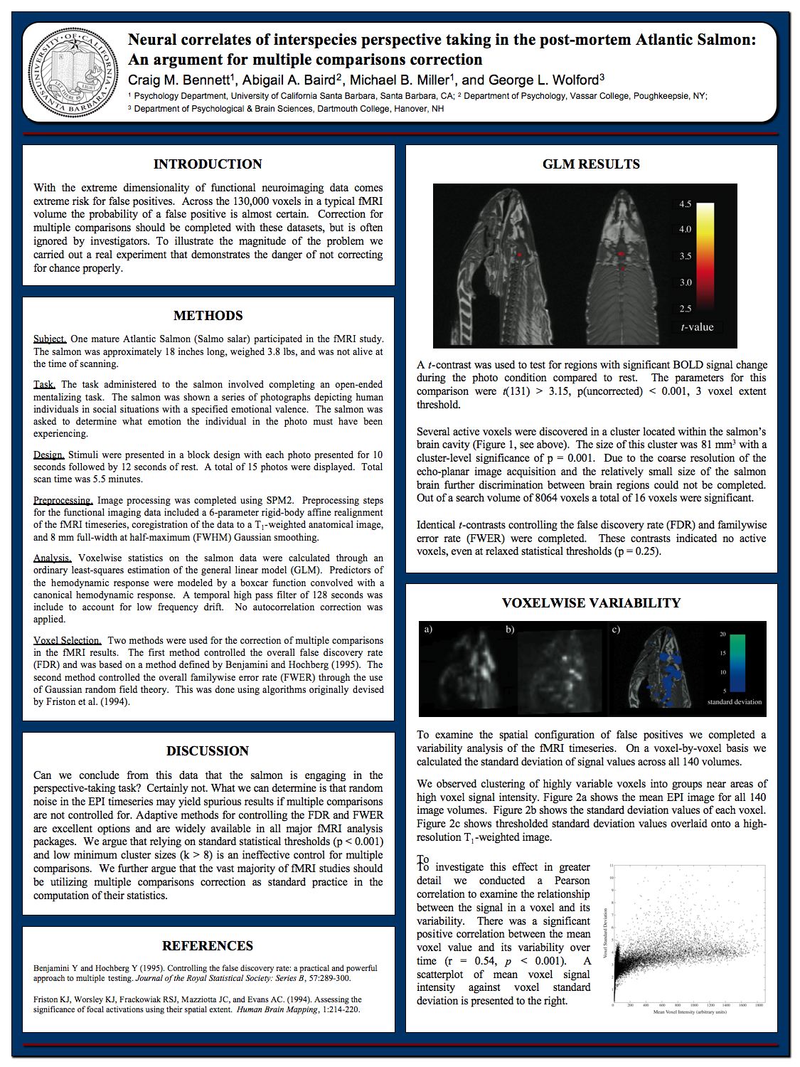
Flaws in brain imaging studies were highlighted by a study that found "correlations of consciousness" by using an fMRI brain scan on a dead salmon. See here for an image summarizing the study.  The dead salmon study highlighted a problem called the multiple comparisons problem. This is the problem that the more comparisons you make between some region of the brain and an average, the more likely you will be to find a false positive, simply because of chance variations. A typical brain scan study will make many such comparisons, and in such a study there is a high chance of false positives. 

Considering the question of “How Much of the Neuroimaging Literature Should We Discard?” a PhD and lab director states, “Personally I’d say I don’t really believe about 95% of what gets published...I think claims of 'selective' activation are almost without exception completely baseless ”  This link says that a study, "published open-access in the Proceedings of the National Academy of Sciences, suggests that the methods used in fMRI research can create the illusion of brain activity where there is none—up to 70% of the time."

A new study has raised additional concerns about the use of brain imaging in neuroscience.  The study was announced in a Duke University press release entitled, "Studies of Brain Activity Aren't as Useful as Scientists Thought."  The study discusses a meta-analysis which looked at the question of how reliably there occurs a region of higher brain activation, in cases when a particular subject had his brain scanned at two different times. 

What neuroscientists would like for there to be is a tendency to get the same result in two different scans of a person's brain taken on two different days, when the person was engaged in the same activity or exposed to the same stimulus.  But that doesn't happen.  We read the following in the press release, which quotes Ahmad R. Hariri:

"Hariri said the researchers recognized that 'the correlation between one scan and a second is not even fair, it’s poor.'...For six out of seven measures of brain function, the correlation between tests taken about four months apart with the same person was weak....Again, they found poor correlation from one test to the next in an individual. The bottom line is that task-based fMRI in its current form can’t tell you what an individual’s brain activation will look like from one test to the next, Hariri said....'We can’t continue with the same old ‘"hot spot" research,' Hariri said. “We could scan the same 1,300 undergrads again and we wouldn’t see the same patterns for each of them.”

The press release is talking about a scientific study by Hariri and others that can be read here or here.  The study is entitled, "What is the test-retest reliability of common task-fMRI measures? New empirical evidence and a meta-analysis." The study says, "We present converging evidence demonstrating poor reliability of task-fMRI measures...A meta-analysis of 90 experiments (N=1,008) revealed poor overall reliability."

In a neuoscience study, the sample size is how many subjects (animal or human) were tested. Figure 1 of the Hariri study deserves careful attention. It has three graphs comparing the kind of sample sizes we would need to get reliable results in brain imaging studies (ranging from between 100 and 1000) to the median samples size of brain image studies (listed as only 25).  This highlights a problem that I have many times written about: that the sample sizes used in neuroscience studies are typically way too small to produce reliable results. As it happens, the problem is even worse than depicted in Figure 1, because the median sample size of a neuroscience study is actually much less than 25. According to the paper here,  "Highly cited experimental and clinical fMRI studies had similar median sample sizes (medians in single group studies: 12 and 14.5; median group sizes in multiple group studies: 11 and 12.5)."

Neuroscientists have known about this shortcoming for years. It has been pointed out many times that the sample sizes used in neuroscience studies are typically way too small for reliable results. But our neuroscientists keep grinding out countless studies with too small a statistical power. In the prevailing culture of academia, you are rewarded for the number of papers published with your name on it, and not too much attention is paid to the reliability of such studies. So if you are a professor with a budget that is sufficient to fund either 100 fMRI scans on 100 subjects in a single study of relatively high reliability, or 10 little low-reliability studies with only 10 subjects each, the prevailing rewards system in academia makes it a better career move for you to do 10 unreliable studies resulting in 10 separate papers rather than a single study resulting in a single paper.

Figure 5 of the Hariri study is also interesting. It rates reliability in various tests of mental activity while subjects had their brains scanned at two different times.  There's data for a single task involving memory, which failed to reach a reliability of either "excellent" or "good."  This task involved a retest of only 20 different subjects. On the left of the figure, we have results for an Executive Function (EF)  test tried twice on 45 subjects, and a "relational" test tried twice on 45 subjects. The relational test is discussed here.  In the test you have to look at some visual figures, and mentally discern whether the type of transformation (either shape or texture) that occurred in a first row of figures was the same transformation used in the second row of figures.

So we have here the interesting case of two thinking tasks applied to 45 subjects on two different days, while their brains were scanned. This makes a better-than-average test of whether some brain region should reliably be activated more strongly during thinking.

The result was actually a flop and a fail for the hypothesis that your brain produces thinking.  In the Executive Function test (corresponding to the third column of circles shown below), none of the 8 brain regions examined produced a greater activation that appeared to an extent that was either Excellent, Good, or Fair.  In the relational test (corresponding to the fifth column of circles shown below), none of the 8 brain regions examined produced a greater activation that appeared to an extent that was either Excellent, Good, or Fair.  The figure is shown below:

brains do not cause thinking
Figure 5 of the Hariri study (link)

The brain regions used in the tests graphed above were not random brain regions, but were typically the regions thought most likely to produce a correlation.

Such results are quite consistent with the claim I have long made on this blog that the brain is not the source of the human mind, and is not the source of human thinking.

Postscript: The study "Reliability and stability challenges in ABCD task fMRI data" (by James T. Kennedy and others) finds results like that of the Hariri study, but much more damaging to claims of reliability in brain scanning studies. The Kennedy study examined how well retest scans of thousands of individuals matched results found in the original scans done days or months earlier.  Referring to a numerical measure in which any result less than .4 means "poor" with little replication between different scans of the same subject, we read the following ridiculously bad results:

"Reliability and stability (quantified as the ratio of non-scanner related stable variance to all variances) was poor in virtually all brain regions, with an average value of 0.088 and 0.072 for short term (within-session) reliability and long-term (between-session) stability, respectively, in regions of interest (ROIs) historically-recruited by the tasks. Only one reliability or stability value in ROIs exceeded the ‘poor’ cut-off of 0.4, and in fact rarely exceeded 0.2 (only 4.9%)."

What this means is the brain scans showed virtually no replication beween scans done on the same individual doing the same task on different days. The result is consistent with the idea that brain scans merely pick up random fluctuations, without revealing any evidence for claims that brains make minds.  

Friday, June 5, 2020

Global Workspace Theory Sure Isn't an Explanation for Consciousness

Neuroscientists have no credible explanations for the most important mental phenomena such as consciousness and memory. All that scientists have in this regard are some far-fetched speculations or weak theories that don't hold up to scrutiny.  Supposedly the two most popular theories of consciousness proposed by scientists are one theory called integrated information theory and another theory called global workspace theory. You can read here why integrated information theory does not work as a credible theory of consciousness.  Global workspace theory isn't any better.

The wikipedia article on global workspace theory starts out by explaining it this way:

"GWT can be explained in terms of a 'theater metaphor.' In the 'theater of consciousness'  a 'spotlight of selective attention' shines a bright spot on stage. The bright spot reveals the contents of consciousness, actors moving in and out, making speeches or interacting with each other. The audience is not lit up—it is in the dark (i.e., unconscious) watching the play. Behind the scenes, also in the dark, are the director (executive processes), stage hands, script writers, scene designers and the like. They shape the visible activities in the bright spot, but are themselves invisible."

As a causal explanation for why a brain might be able to produce understanding or consciousness, this is a complete failure, as it does not refer to anything in the brain, but refers to some theater. At most it is some metaphor merely describing selective attention, but selective attention (or mental focus) is merely an aspect of understanding once it exists, not an explanation of consciousness or understanding.  You can't spotlight your way to consciousness. Also, there's nothing in the brain that actually corresponds physically to a spotlight. When you're thinking about something, it is not at all true that some particular region of your brain lights up like some area under a spotlight, contrary to the misleading statements and misleading visuals often given on this. Actual signal strength differences (typically far less than 1%) are no greater than we would expect from random variations.  

In an interview in Scientific American, Bernard Baars attempts to explain global workspace theory, but fails rather miserably to give a coherent explanation of how global workspace theory is anything like a theory explaining consciousness.   He is asked by the interviewer, "What is global workspace theory?" What we then get from Baars is  an answer that kind of wanders around all over the place for 11 paragraphs without giving much of any answer that anyone will be able to grasp. 

There is some mention of some swarm computing setup: "If you put a hundred crummy algorithms together and let them share hypotheses and vote on the most popular one, it turns out that very inadequate algorithms could jointly solve problems that no single one could solve." There is entirely irrelevant for any explanation of consciousness or understanding, because particular areas of the brain are not like little micro-processors running software code. There is nothing like software code that runs anywhere in the brain. 

Baar's rambling and muddled answer to the question ends like this:

"Part IV of my latest book On Consciousness: Science & Subjectivity develops GW dynamics, suggesting that conscious experiences reflect a flexible 'binding and broadcasting' function in the brain, which is able to mobilize a large, distributed collection of specialized cortical networks and processes that are not conscious by themselves. Note that the 'broadcast' phase proposed by the theory should evoke widespread adaptation, for the same reason that a fire alarm should evoke widespread responding, because the specific needs for task-relevant responders cannot be completely known ahead of time. General alarms are interpreted according to local conditions. A brain-based GW interacts with an 'audience' of highly distributed, specialized knowledge sources, which interpret the global signal in terms of local knowledge (Baars, 1988). The global signal triggers reentrant signaling, resonance is the typical activity of the cortex."
Baar's scrambled 11-paragraph answer is a complete failure as an attempt to explain how a brain could produce consciousness or understanding. Electrical signals travel around in the brain, but there is nothing like a broadcast in the brain that could explain consciousness or understanding.  And it's rather silly to be trying to use fire alarms as part of an attempt to explain consciousness or understanding. 
To understand how impotent the idea of broadcasting is to explain consciousness or understanding, let's consider the city I grew up in. When I was a boy there were in my city several very high broadcast towers that broadcasted TV signals and radio signals. Almost every house in the city had an old-fashioned TV that picked up these TV signals, and also an old-fashioned radio that picked up the old-fashioned radio signals. But none of this huge amount of broadcasting and broadcast reception resulted in the slightest bit of consciousness in any of the antennas, the television sets or the radios.  The idea of broadcasting is worthless in explaining consciousness. 
We cannot at all explain consciousness by saying that it adds up from the activity of a bunch of networks that "are not conscious by themselves."  There is no reason why the activity of a bunch of unconscious networks should add up to be a conscious reality, any more than having a house made of bricks should add up to be a wooden house.

The reality in the brain is that there are billions of cells that each emits electrical signals.  A rough analogy might be a packed stadium with 80,000 people who are each making noise during a football game.  But still you have a unified self and a unified stream of thought from a mind. There's not the slightest reason why that would emerge from the activity of billions of individual neurons, just as there's not the slightest reason why a single paragraph of speech would ever flow from the lips of 80,000 people in a stadium.

A broadcast is a stream of tokens that can give information to an agent capable of understanding who is listening to such a broadcast. But a broadcast does nothing to ever produce such an agent of understanding.  The flow of tokens during a broadcast is rather like the stream of bullets from a machine gun.  Thinking that you can broadcast your way to consciousness is as silly as thinking that you can machine-gun your way to consciousness.

Narrating an achievement legend that is groundless (something very common these days in academia), Baars makes these mostly false claims:
"Our individuality is a function of the cortex, which is now proven by brain studies to be 'the organ of consciousness.' Wilder Penfield discovered that in 1934 via open-brain surgeries in fully awake patients, who were able to talk with him and gesture."
The brain is an organ, and the cortex is not an organ, but only a small fraction of an organ. So calling the cortex "the organ of consciousness" is nonsense.  There are no brain studies showing that the cortex produces consciousness. To the contrary, we know that after hemispherectomy operations in which half of the cortex (and half of the brain) is removed and discarded, to stop very bad seizures,  people are just as conscious and just as intelligent as they were before such an operation.  And we also know from the studies of people like physician John Lorber that people have existed with very good consciousness and above-average intelligence, even though they had brains and cortexes that were almost entirely destroyed by disease. Such medical case histories debunk claims that the cortex is the source of consciousness. Of course, an operation by Penfield in which people can talk and gesture during brain surgery does absolutely nothing to establish that the brain or the cortex is the source of consciousness.  So it is wrong for Baars to be citing such a thing as evidence that Penfield discovered that consciousness comes from the cortex.

Baars has tried to suggest the idea that consciousness comes from a broadcasting of something from the cortex. But the cortex of the brain is an actually an extremely bad broadcaster.  Electrical signals in the cortex travel from one neuron to another with a very low reliability.  It has been estimated that the chance of an action potential traveling between two adjacent neurons in the cortex is below 50%, and as low as 10%.  A scientific paper says, "In the cortex, individual synapses seem to be extremely unreliable: the probability of transmitter release in response to a single action potential can be as low as 0.1 or lower." It's implausible to be saying that cortex cells that are such bad and unreliable information transmitters (such bad broadcasters) are somehow giving rise to consciousness through some kind of broadcasting effect. 
Baars has some book describing his ideas on this topic.  But I see no reason why anyone should buy such a book, because nothing that Baars states in his Scientific American interview should give us any confidence that he has any substantive explanation for how a brain could produce consciousness, thinking or understanding.  When asked about the "hard problem of consciousness" he states there is no evidence for it, which makes no sense, and is like saying there is no evidence for the problem of the origin of language or the problem of the origin of life.  
Postscript: Some have claimed that Wilder Penfield's experiments show memories are stored in brains, but such claims are unwarranted. Penfield's experiments are described in the book The Human Mind Explained (page 132):

"When this area [the temporal lobe] was stimulated, 40 out of about 1000 patients reported vivid flashbacks -- a fragment of a tune, a child calling, being in a room. All were marked by a dreamlike quality with no sense of time or location. He believed that this was evidence that our memories are stored in one place in a complete and recoverable form. Later researchers pointed out, however, that few of the patients recalled actual memories."  

page online says Penfield "did not provide support for the claim that what was elicited was actually a memory and not a hallucination, fantasy or confabulation." A book has a page dealing with Penfield's brain stimulation experiments, and quotes some expert saying such stimulation does not directly activate memory traces.  The same page refers to "dreamlike mentation" produced by such stimulation, "in the Lewis Carroll mode," such as someone seeing pigs walking around upright like people.