Monday, September 30, 2024

Brains Show No Physical Signs of Reading or Retrieval When You Recall Something

There are three neutral words you can use to describe remembering:

(1) You can use the neutral word "remembering," which does not imply anything physical occurring. 

(2) You can use the neutral word "recall," which does not imply anything physical occurring.  That's a good word to use in describing a case when someone recites facts after hearing a single word or name, or answers a question. 

(3) You can use the neutral word "recognition," which does not imply anything physical occurring. That's a good word to use to describe cases in which you see a photo or face, and identify a person, place or object. 

There is another common word used to refer to remembering: the word "retrieval." The word implies some act involving going and getting something, and bringing it from one position to another. For example, the word "retrieval" is used for what goes on when a dog owner plays the game of "fetch," by tossing a stick, and then watching his dog go get the stick and bring it back to the owner.  When I search for the definition of "retrieval" the first definition I get is: "the process of getting something back from somewhere." The Cambridge Dictionary defines "retrieval" as "the process of finding and bringing back something."

A reality with very big implications is this: when a computer retrieves some information, there can be definite physical signs of both reading and retrieving; but when a human being remembers something, there is no physical sign in the brain of any such thing as either reading or retrieval. 

To help explain this contrast, let us first look at a simple case of information retrieval using a somewhat old-fashioned desktop computer of the type that was predominant around the year 2010.  We may consider a very simple case of someone at a keyboard typing something that commands the computer to display some image stored on the computer. Normally a mouse would be used as part of the retrieval, but the same thing can be done with the keyboard only. For example, if  my computer shows a Windows folder on the screen, I can use keys such as the arrow keys to navigate to a particular image file, and then press the Enter key to cause some particular image file to be displayed on the monitor. 

The visual below illustrates part of what is going on. The red arrows signify data or signals that are passing around from one part of the system to another. 

computer retrieval of information

1. First, an electronic signal is sent from the keyboard to the computer, a signal corresponding to a request that a particular image file be displayed. 

2. Then the image file is read from the hard drive of the computer, by an act that requires that the read/write head of a hard drive lines up with a particular spot on the hard drive.

3. Then an electronic stream of bits passes from the computer to the monitor, causing the image to be displayed on the screen. 

This involves real reading, and real retrieval. Just like a set of human eyes that focuses on some particular line of text while it is reading, the read/write head of the hard drive focuses on one tiny spot of the spinning hard drive disk, to do some reading. Real retrieval is going on because some chunk of data is found, and is also copied from its source location (in the computer) to a destination location (the monitor).  The only quibble you could make about the use of the word "retrieval" to describe this is that rather than the original data being moved from its original location to its target location, the data is actually copied, leaving the source data unchanged. The resulting act is kind of like what would happen if you retrieved a book from a shelf, but magically got a copy of the book when you touched the original, leaving the original on the shelf. 

Now, let us consider the interesting question: is there any evidence of a physical process anything like this in the brain when someone remembers something? There is not. 

We can imagine some extraterrestrial being with a brain that might act in a manner similar to the computer just described. That being's brain might work such as this:

(1) The extraterrestrial's brain might have some "attention center" part that is inactive whenever the extraterrestrial being is not thinking or remembering, but active when the extraterrestrial being is thinking or remembering.

(2) The extraterrestrial's brain might have some "roving cursor" feature that served as a memory reading unit. 

(3) When the extraterrestrial remembered something, the "roving cursor" unit might move to some particular part of the brain where the information was stored, and read from that part.  Then the "roving cursor" might bring back that information from that spot to the "attention center" of the extraterrestrial's brain, causing the extraterrestrial being to have a thought corresponding to the memory. 

But the human brain seems to have nothing whatsoever like anything I have imagined above. In particular:

(1) There is no sign of anything like some "attention center" in the brain which is inactive during mental inactivity and only active during thinking or retrieval. 

(2) There is no mobile unit whatsoever in the brain that moves around to cause some act of memory retrieval. 

(3) There are in the brain no indexes, sorting or addresses that might allow some item of information to be instantly found, such as occurs when you are asked a question of who was some historical figure, and you immediately state a few sentences describing that figure. 

The four pillars of instant knowledge retrieval in physical systems

(4) Other than the mechanism for reading genetic information that is not learned information, a mechanism existing throughout the body, there is no known mechanism by which an act of reading stored information can occur in the brain.  The brain has nothing like a read/write head in a computer. While the eye has a focus mechanism allowing it to focus on some particular spot in space, the brain has nothing like some focus mechanism allowing it to focus on some particular group of cells in the brain. 

(5) When someone remembers something, there is no evidence of some transfer of data from a storage place to some "attention place" when someone remembers something. When someone remembers something, there is no evidence of any transfer of data corresponding to the memory recall.  Chemicals are constantly traveling around the brain, as are changes in electrical charge; and at any second a large fraction of all neurons fire.  There is no evidence that such activity increases when someone remembers something. A web page states, "Our best guess is that an average neuron in the human brain transmits a spike about 0.1-2 times per second." So a person wishing to claim that nerve impulses (action potentials) travel from one area of the brain to another when you remember will not be telling an outright lie. But if he merely says that, he is telling a half truth. The fact is that if we compare a moving bubble to a nerve impulse, the brain is like a  pot of boiling water in which bubbles are constantly traveling around all over the place. 

The visual below schematically diagrams the relation between neurons and action potentials. We see what represents a group of neurons, with individual neurons represented as black dots. The red arrows represent electrical charges or chemicals that are travelling around between neurons. Since a neuron fires randomly about once a second, we have a "signal traffic" arrangement that is random.  

brain signal traffic

The diagram above represents just a billionth of the neurons in the brain. With such a situation, there is no impression of data traveling from one place to another in any organized way when memory recall occurs. What we have all over the brain is electrical and chemical noise. 

There is no way of tracking all of these electrical and chemical impulses going around between neurons. There is no way to eavesdrop on the traffic passing around. The idea of doing such a thing is as impractical as listening to all the notes of the birds singing in New York City. What scientists can do is to scan brains using fMRI scanners, and look for differences between mental rest activity and some type of cognitive activity. Such scans show differences in different parts of the brain no greater than about 1 part in 200 from one area to another.  We would expect such differences from mere chance variations. Such differences are not any evidence that memory retrieval occurs by brain activity. 

Besides failing to find any readable memories from a microscopic examination of brain tissue, scientists are unable to "eavesdrop" on a brain or "wiretap" a brain to listen on something like a memory retrieval, and to figure out what a person was recalling from activity in his brain. Nor has anyone ever produced robust evidence of any type of increased electrical transmission or signal transmission occurring in a brain when a human recalls something.  Any claim to have produced evidence of such a thing will be another example of the Questionable Research Practices that today's experimental neuroscientists are so massively guilty of, and will also probably be an example of the pareidolia so commonly occurring among such neuroscientists. Pareidolia typically occurs when someone examines a large, ambiguous, rapidly fluctuating data set, and see whatever he hopes to see (like someone scanning the clouds looking for faces or animal shapes). 

There is no retrieval-related arrow diagram we can make for the brain corresponding to the retrieval-related arrow diagram above showing what goes on when you request the retrieval of an image from your computer. Brains show no physical signs of reading or retrieval when you lie with your eyes closed and recall something. The physical characteristics of the brain and the flow of energy and matter within the brain is perfectly compatible with the belief that the brain is not the storage place of human memories, and that memory recall does not occur through brain activity. 

Monday, September 23, 2024

They Mapped 150 Million Synapses, and Found No Sign of Stored Memories

A recent press account states this:

" Researchers have reconstructed a minuscule piece of the human brain down to the level of individual synapses, representing a giant leap forward for brain science. And we're not just talking about a few neurons here. This millimeter-sized cube contains a staggering 57,000 cells, 230 millimeters of tiny blood vessels, and nearly 150 million synaptic connections, all mapped out in glorious 3D detail."

We are told that the bit of brain tissue mapped was taken from an epilepsy patient, presumably in one of those operations in which brain tissue is removed to help prevent seizures. The brain tissue was divided into 5000 different sections, and then thoroughly photographed with an electron microscope. 

A web site allows access to some of the resulting photos and 3D models, and a scientific paper describing the data. What is cool is that upon looking at one of the photos, you can click with your mouse, and then drag around the image, which will rotate in 3D.

The page here allows you to see some neurons and their connections, and each neuron and its connections is shown in a separate color:

neurons

At the page here you can see a view of some dendrites and their little bumps called dendritic spines. Below is what you first see. By clicking on the right image, you can drag around in 3D the right part. 

dendritic spines

Dendritic spines are the little bumps we see above

We can see from the scales at the bottom how very small are the structures shown. The dendritic spines shown above have a size of only about 1 micrometer, a millionth of a meter. The symbol for a micrometer is "ยต."

Using the page here, you can see a synaptic gap between two connected synapses. The orange and green units on the left are synaptic boutons that correspond to the similar-looking orange and green units on the right. The synaptic clefts are about 300 to 700 nanometers wide. A nanometer is a billionth of a meter, or a thousandth of a micrometer. The symbol for a nanometer is nm. 

synaptic cleft

The investigation of this neural tissue is described in the paper here, entitled "A connectomic study of a petascale fragment of human cerebral cortex." It is interesting to see the discrepancy between the paper's lip-service to neuroscientist dogmas about a brain storage of memory, and the complete failure to supply any observations supporting such dogmas. 

"The most functionally significant aspect of cortical tissue is the synaptic connectivity that allows neurons to send and receive signals to and from other neurons. This wiring diagram is likely central to the way human brains store memory and give rise to behavior....Given the far greater variability in human experience, behavior, memory and genetics, and the fact that humans and other vertebrates have pools of identified neurons classes rather than individual identified neuron types, it will no doubt be challenging to compare neural circuits between brains....Even if the circuits differ in their particulars, it is possible that a metalogic for memory can be uncovered by looking at enough data, maybe in the future field of 'engramics.' To be sure, approaches to the profound questions of uncovering the meaning in neural circuit connectivity data are in their infancy, but it would seem to us that perhaps the best stimulus for making progress will be an abundance of actual data -- this petascale dataset is a start."

Those are all of the references the paper makes to memory, except for a reference to computer memory. The claim that studies looking for evidence of memory storage in brain tissue are in their infancy is incorrect, as is the claim that this study "is a start."  As I document in my post "They Stored and Studied Thousands of Brains, But Still Failed to Show Brains Store Memories," studies like this have been going on for decades.  And none of them have produced any evidence that memories are stored in brains. 

Could it be that the dendritic spines like the ones we see above are some kind of information representation system? There's no chance of that. For one thing, such dendritic spines lack any regularity such as you might have in an information representation system.  Dendritic spines appear with shapes and numbers as random as the buds coming from trees branches. Just as no one analyzing the buds coming from tree branches can detect any kind of code in which some symbolic information is being stored, no one analyzing the dendritic spines coming from dendrites can detect any kind of code in which symbolic information might be stored.  Just as the buds coming from tree branches have none of the regularity that we might find if symbolic information was being stored, the dendritic spines coming from dendrites have none of the regularity that we might find if symbolic information is being scored. And just as there is in nature no such thing as a tree branch bud reader, there is in the brain no such thing as a dendritic spine reader. And just as the buds coming out of tree branches don't last very long (lasting much less than a year), the dendritic spines coming out of dendrites don't last very long. For details see my post "Imaging of Dendritic Spines Hint That Brains Are Too Unstable to Store Memories for Decades."  The average lifetime of a dendritic spine is only about 120 days. 

Another example of the empty hand-waving of neuroscientists in regard to memory can be found in the paper here, entitled "Why not connectomics?" We have this example of conceptually empty hand-waving about memory storage:

"Brains can encode experiences and learned skills in a form that persists for decades or longer. The physical instantiation of such stable traces of activity is not known, but it seems likely to us that they are embodied in the same way intrinsic behaviors (such as reflexes) are: that is, in the specific pattern of connections between nerve cells. In this view, experience alters connections between nerve cells to record a memory for later recall. Both the sensory experience that lays down a memory and its later recall are indeed trains of action potentials, but in-between, and persisting for long periods, is a stable physical structural entity that holds that memory. In this sense, a map of all the things the brain has put to memory is found in the structure—the connectional map."

The first sentence is groundless dogma. There is no evidence that brains "can encode experiences and learned skills in a form that persists for decades or longer."  There is merely the fact that humans can have experiences and learn skills that they remember for decades.  The beginning of the second sentence is a confession that there is no understanding of how such a brain storage of memories can happen. The authors confess that "the physical instantiation of such stable traces of activity is not known,"  The claim that memories are stored by "the specific pattern of connections between nerve cells" is empty hand-waving, and the speculation stated is unbelievable. No one who has ever studied the connections between nerve cells (neurons) has ever seen anything like some symbolic pattern that could encode a record of human experiences or human learned skills or learned conceptual knowledge such as school learning.  The brain does not have any such thing as a connection pattern reader that could read and interpret such patterns if they existed. 

problems with materialist ideas


Monday, September 16, 2024

No, They Didn't Find in a Brain "3 Copies of Every Memory," and They Never Even Found One

In my post "Why the Academia Cyberspace Profit Complex Keeps Giving Misleading Brain Research Reports" I discussed the economic reasons why we keep getting misleading research about brains, and misleading headlines about brain research. The analysis in that post holds true not just for brain research, but for scientific research in general. We live in an economy in which misleading stories about scientific research and groundless but interesting-sounding scientific speculation are highly incentivized. To give a short synopsis of what I discussed at much greater length in that post, the economic motivations are like this:

(1) Scientists are judged by how many papers they publish and how many citations such papers get.

(2) Because of publication bias (in which papers reporting positive results and particularly interesting-sounding positive results are more likely to be published), scientists are strongly motivated to publish papers claiming positive results and also claiming interesting-sounding results.

(3) Wishing to make themselves appear like sources of important research breakthroughs to help justify their exorbitant tuition, universities are motivated to produce press releases exaggerating the importance of research papers published by their professors.

(4) Since science news is published on web pages with ads that generate revenue for the people running or funding the web pages, with revenue proportional to how interesting-sounding a story is, those running science news web sites or science analysis web sites have an enormous economic motivation to create clickbait headlines that generate higher numbers of page views, and more advertising revenue. Science news sites these days are almost always built in the form of headlines that you must click to read the story, and each time this causes a web page with ads to appear, the people running or funding the site get money from views of the ads displayed on the page you opened up.

The result of all of this is a very wacky world we might call the world of scitainment, to coin a word that combines the words "science" and "entertainment." Scitainment is a part of the internet that blends science and entertainment. Very much of what we read in this strange world of scitainment is true, and very much of it is false. The world of scitainment blends fact and fantasy, always trying its best to produce entertaining stories and clickbait headlines. It's all about luring you in to click on the stories, so that you go to pages that generate ad revenue for the people running the web sites. 

science hype

One of the web sites involved in pushing scitainment is the ad-heavy site www.livescience.com, where we have many science headlines that simply are not true. To give some examples:

  • On the Livescience site we had the utterly untrue headline "Building blocks of life' discovered on Mars in 10 different rock samples." The story discusses some observations of biologically irrelevant chemicals on Mars, none of which are ingredients of life or building blocks on life.  
  • The same Livescience site had an article claiming a woman was hit by a meteorite while drinking coffee outside, although a space.com story tells us no such thing happened. 
  • A story at the LiveScience site was entitled " 'This might be the seeds of life': Organic matter found on asteroid Ryugu could explain where life on Earth came from." The story was rubbish for several reasons: (1) Scientists do not believe that life ever existed on the asteroid  Ryugu or on any other asteroid. (2) There is no scientific concept of any such thing as a "seed of life," in the sense of something causing life to arise from non-life (with the exception of plant seeds, and plant seeds were not found on Ryugu).  (3) No actual components of life were found on the asteroid Ryugu, and most organic molecules are not components of life. 

  • Another story at the LiveScience site referred to a claimed discovery of the simplest amino acid (uracil) on an asteroid, in the faintest trace amount of only 13 parts per billion. The headline at the LiveScience site made the very untrue claim that this "could explain the origin of life." Living things require twenty types of amino acids, which must be massively arranged in very specially ordered arrangements to make many types of the very hard-to-achieve molecules called proteins.  The discovery of one type of amino acid in the faintest trace amounts no more explains the origin of life than the discovery of a twig on the ground (making the letter "I") explains the origin of books consisting of vey much well-constructed prose. 

  • Another article on the LiveScience site was devoted to selling the groundless idea that there is a "dark mirror" universe inside ours. 

  • Another article on the LiveScience site had the nutty title "The 1st life in the universe could have formed seconds after the Big Bang."  Anyone familiar with the incredibly high temperatures and density at such a time (preventing all chemistry and even the existence of atoms) should understand how crazy such a claim is. 

  • Another article on the LiveScience site had the phony title "Here's what we learned about aliens in 2020," a reference to extraterrestrials. Of course, we did not learn anything about extraterrestrials in that year. 

  • Another article on the LiveScience site had the phony title "These weird lumps of 'inflatons' could be the very first structures in the universe."  We saw a visual of some strange structure that looked like a planetary nebula. The caption read, "Shown here, one of the dense clumps of inflatons that emerged during the inflation phase of the Big Bang, in the infant universe."  The caption led the reader to believe he was looking at some photo of something in space.  But the photo was not a photo of anything observed in space.  It was merely a photo of some junk generated by an entirely speculative computer program. No actual "inflatons" have ever been observed, and the program was based on one of the innumerable speculative models of the unproven cosmic inflation theory.


As the examples above show, you should not assume a claim is true merely because you read a headline suggesting it is true at the LiveScience site at www.livescience.com.   The latest example of a misleading headline at the site is an article with the groundless headline "The brain stores at least 3 copies of every memory." Human beings recall things, but no scientist has ever discovered even one memory in a brain. 

You can pretty much figure out that the story is baloney the moment you read that the research discussed is merely research based on mice rather than humans.  Letting our imaginations run wild, we can imagine some investigator of human brain tissue confirming the claim that brains keep three different copies of each memory. For example, an investigator might keep scanning the brain of a dead person, and then announce something like, "I found the words 'the battle of Hastings occurred in 1066' in three different spots of the brain." But we can imagine no possible observations of mice brains that would ever justify the claim that a memory was stored in a mouse brain.  For example, a researcher could never announce that he found the words "mouse traps are dangerous" in some part of a mouse brain, simply because mice don't use language. 

Misspeaking both in its headline and in its text, the article says, "The scientists found that, in rodents, the brain stores at least three copies of a given memory, encoding it in multiple places in the organ." No, scientists found no such thing. The article refers to the junk-science paper "Divergent recruitment of developmentally defined neuronal ensembles supports memory dynamics." It is true that in the abstract of the paper the authors claim " we discovered that memory encoding resulted in the concurrent establishment of multiple memory traces in the mouse hippocampus."  But because the authors used very bad research practices, they provided not the slightest bit of robust evidence for such a claim. 

The paper is behind a paywall, but anyone can read a preprint of the paper that allows us to see the Questionable Research Practices that were used.  The defects are as follows:

(1) The study group sizes were way-too-small, consisting of groups such as only 4 mice or only 5 mice or only 8 mice or only 10 mice. No one should take seriously any experimental rodent research study using fewer than 15 rodents in each study group, and for most effect sizes a larger study group size such as 30 mice is needed. The authors would have discovered the inadequacy of their study group sizes if they had done a sample size calculation like good scientists, but they failed to do that. The paper "Prevalence of Mixed-methods Sampling Designs in Social Science Research" has a Table 2 giving recommendations for minimum study group sizes for different types of research. The minimum number of subjects for an experimental study is 21 subjects per study group. 

minimum sample sizes


(2) We hear no discussion of the following of a detailed blinding protocol, something that would need to exist for a study like this to be taken seriously. The only mention of a blinding procedure is the mere remark that "To reduce potential bias in the analysis, the researcher conducting the analysis was blind to the experimental
group to which animals belonged until after the data analysis was completed."  When you are using very small study group sizes such as only 4 mice or only 8 mice, it is typically the case that mice can be recognized visually, meaning a researcher can tell things he was not told, such as whether a mouse was in a control group.  Serious use of a blinding protocol requires a careful protocol that would require at least a long paragraph to state, and we have no evidence of such a thing in this paper. 

(3) The experiment was thoroughly entangled with the use of a worthless technique for measuring memory recall in mice, the defective technique of trying to judge "freezing behavior" in mice.  The preprint paper uses the word "freezing" 77 times, to show how the experiments were thoroughly dependent upon the use of such a technique. All experimental neuroscience papers depending on such judgments of "freezing behavior" are junk science papers. 

"Freezing behavior" judgments work like this:

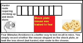
(1) A rodent is trained to fear some particular stimulus, such as a red-colored shock plate in his cage. 

(2)  At some later time (maybe days later) the same rodent is placed in a cage that has the stimulus that previously provoked fear (such as the shock plate). 

(3) Someone (or perhaps some software) attempts to judge what percent of a certain length of time (such as 30 seconds or 60 seconds) the rodent is immobile after being placed in the cage. Immobility of the rodent is interpreted as "freezing behavior" in which the rodent is "frozen in fear" because it remembered the fear-causing stimulus such as the shock plate. The percentage of time the rodent is immobile is interpreted as a measurement of how strongly the rodent remembers the fear stimulus. 

This is a ridiculously subjective and inaccurate way of measuring whether a rodent remembers the fear stimulus. There are numerous problems with this technique:

(1) There are two contradictory ways in which a rodent might physically respond after seeing something associated with fear: a flight response (in which the rodent attempts to escape) and a freezing response (in which the rodent freezes, not moving). It is all but impossible to disentangle which response is displayed when the rodent is presented with a fear stimulus. A rodent who remembers a fear stimulus might move around trying to escape the feared stimulus. But under the "freezing behavior" method, such movement would not be recorded as memory of the feared stimulus, even though the fear stimulus was recalled. 

(2) Rodents often have hard-to-judge movement behavior that neither seems like immobility nor fleeing behavior, and it is subjective and unreliable to judge whether such movement is or is not "freezing behavior" or immobility. 

(3) Movement of a rodent in a cage may be largely random, and not a good indication of whether the rodent is afraid and whether the rodent is recalling some fear stimulus. 

(4) Rodents encountering a fear-provoking stimulus in human homes (such as a mouse hearing a human shriek) almost never display freezing behavior, and much more commonly display fleeing behavior. I lived in a New York City apartment for many years in which I would suddenly encounter mice, maybe about 10 times a year. I never once saw a mouse freeze when I shrieked upon seeing it, but invariably saw the mouse flee. 

(5) Freezing behavior in a rodent may  last for a mere instant, as in humans. So it may be extremely fallacious to do something such as trying to observe 30 seconds or 60 seconds of rodent movement or non-movement, and try to judge whether fear or recall occurred  by judging a "freezing percentage" over such an interval. Almost all of that time may be random behavior having nothing to do with fear in the rodent or memory recall in the rodent. 

For experiments not involving recall of a fearful stimulus, the Morris Water Maze test can be used to reliably measure recall in rodents. There are two reliable ways to measure fear recall in rodents. The first is to measure heart rate, which very dramatically spikes in rodents when they are afraid. The second is to measure an avoidance of a fearful stimulus.  The simple technique is illustrated in the visual below:

But instead of using such reliable techniques, our neuroscientists continue to use the very unreliable technique of trying to judge recall of fear-related memories in animals by making subjective judgments of "freezing behavior." Why would they continue to use so stupid and unreliable a technique? I can think of two reasons:

(1) Neuroscientists are People of Custom just like Roman Catholic priests are People of Custom. So neuroscientists may keep using some very old and ineffective technique as a matter of "clinging to the old custom," rather like the way Roman Catholic priests kept reciting the Mass in Latin very long after almost no one understood Latin. 

(2) Neuroscientists may prefer to use an unreliable technique for measuring fear-related memory recall in rodents, because using that bad technique increases the chance of them producing research papers that report invalid but interesting-sounding results consistent with "brains store memories" dogmas.  Similarly, if a researcher uses an unreliable technique for detecting heat traces in clouds, it will increase the chance that he can end up with some paper claiming to show heat blips in clouds that he may claim as evidence for extraterrestrial spaceships in the sky. The unreliable measurement technique is the best friend of the person trying to support untrue claims. 

Thoroughly dependent on a bad measurement technique for judging whether rodents recalled a fearful stimulus, and also involving way-too-small study group sizes such as only 4 or 5 rodents, the low-quality science paper "Divergent recruitment of developmentally defined neuronal ensembles supports memory dynamics" has provided zero robust evidence that there is a copy of a memory in any brain. No such robust evidence has ever been provided by neuroscientists. As discussed in my post here, the quickly-preserved brains of thousands of people have been thoroughly studied by different "brain bank" projects, and by microscopic examination no one ever found the slightest evidence of a memory stored in a brain. Never through microscopic examination of a brain has even a single piece of information as small and humble as "birds fly" or "dogs bark" or "Earth has a moon" ever been found. nor has anyone ever found in any brain by microscopic examination even the crudest or blurriest  image of anything anyone saw. 

typical neuroscience press release
Click on the image to read it better

typical neuroscience paper

Postscript:  When "freezing behavior" judgments are made, there are no standards in regard to how long a length of time an animal should be observed when recording a "freezing percentage"  (a percentage of time the animal was immobile). An experimenter can choose any length of time between 30 seconds and five minutes or more (even though it is senseless to assume rodents might "freeze in fear" for as long as a minute).  Neuroscience experiments typically fail to pre-register experimental methods, leaving experimenters free to make analysis choices "on the fly," after they have gathered data. So you can imagine how things might work. An experimenter might judge how much movement occurred during five minutes or ten minutes after a rodent was exposed to a fear stimulus. If a desired above-average amount of immobility (or a desired below-average amount of immobility) occurred over 30 seconds, then 30 seconds would be chosen as the interval to be used for a "freezing percentage" graph. Otherwise,  if a desired above-average amount of immobility (or a desired below-average amount of immobility) occurred over 60 seconds, then 60 seconds would be chosen as the interval to be used for a "freezing percentage" graph. Otherwise,  if a desired above-average amount of immobility (or a desired below-average amount of immobility) occurred over two minutes, then two minutes would be chosen as the interval to be used for a "freezing percentage" graph. And so on and so forth, up until five minutes or ten minutes. Such shenanigans drastically depart from good, honest, reliable experimental methods. 

The paper discussed above did not pre-register any methods, so after gathering data the experimenters were free to analyze the data in any way they pleased. Some of their "freezing behavior" graphs are made using a time interval of three minutes, and others are made using a time interval of five minutes. Genuine fear-freezing in an animal would be something lasting only a few seconds. The longer the interval of time used as a basis for a "freezing behavior" graph, the more unreliable freezing behavior judgments are as a measurement of fear recall. When an interval of longer than 30 seconds is used as the basis for a "freezing percentage" graph, then you have a particularly unreliable and particularly deplorable use of such a technique; and the longer the time interval is above 30 seconds, the more unreliable and deplorable are claims that such graphs are measurements of how well an animal recalled something. 

Monday, September 9, 2024

Neuroscientists Senselessly Think They Can Perform Innumerable Contortions of Brain Data, and Then Claim a Discovery

Claims by neuroscientists that they have found "representations" in the brain (other than genetic representations) are examples of what very abundantly exists in biology: groundless achievement legends. There is no robust evidence for any such representations. 

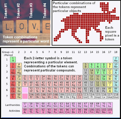
Excluding the genetic information stored in DNA and its genes, there are simply no physical signs of learned information stored in a brain in any kind of organized format that resembles some kind of system of representation. If learned information were stored in a brain, it would tend to have an easily detected hallmark: the hallmark of token repetition.  There would be some system of tokens, each of which would represent something, perhaps a sound or a color pixel or a letter. There would be very many repetitions of different types of symbolic tokens.   Some examples of tokens are given below. Other examples of tokens include nucleotide base pairs (which in particular combinations of 3 base pairs represent particular amino acids), and also coins and bills (some particular combination of coins and bills can represent some particular amount of wealth). 

symbolic tokens

Other than the nucleotide base pair triple combinations that represent mere low-level chemical information such as amino acids, something found in neurons and many other types of cells outside of the brain, there is no sign at all of any repetition of symbolic tokens in the brain. Except for genetic information which is merely low-level chemical information, we can find none of the hallmarks of symbolic information (the repetition of symbolic tokens) inside the brain. No one has ever found anything that looks like traces or remnants of learned information by studying brain tissue. If you cut off some piece of brain tissue when someone dies, and place it under the most powerful electron microscope, you will never find any evidence that such tissue stored information learned during a lifetime, and you will never be able to figure out what a person learned from studying such tissue.  This is one reason why scientists and law enforcement officials never bother to preserve the brains of dead people in hopes of learning something about what such people experienced during their lives, or what they thought or believed, or what deeds they committed.    

But despite their complete failure to find any robust evidence of non-genetic representations in the brain, neuroscientists often make groundless boasts of having discovered representations. What is going on is pareidolia, people reporting seeing something that is not there, after wishfully analyzing large amounts of ambiguous and hazy data. It's like someone eagerly analyzing his toast every day for years, looking for something that looks like the face of Jesus, and eventually reporting he saw something that looked to him like the face of Jesus.  It's also like someone walking in many different forests, eagerly looking for face shapes on trees, and occasionally reporting a success, or like someone scanning the sky, looking for clouds that look like animal shapes.

pareidolia

The latest example of nonsensical neuroscientist pareidolia is to be found in a press release from Columbia University, and the paper that press release describes in very misleading terms.  The press release has the phony headline "
Scientists Capture Clearest Glimpse of How Brain Cells Embody Thought." When you read a headline like that, you should remember a sad truth that has been glaringly obvious for many years now: the fact that university press releases on topics of science are no more trustworthy than corporate PR press releases.  There is the most gigantic amount of lying, hype and misrepresentation in university press releases these days, and such baloney occurs in equal amounts in the press releases of every major university. So don't think for a moment than you can  trust a press release because it came from Harvard or Columbia or Yale or Oxford University. I wish I had a dollar for every bogus press release that has been issued by such institutions. The subtitle of the press release is the 100% untrue claim "Recordings from thousands of neurons reveal how a person’s brain abstractly represents acts of reasoning."

We have an utterly groundless claim by a neuroscientist that he and his colleagues found a “uniquely revealing dataset that is letting us for the first time monitor how the brain’s cells represent a learning process critical for inferential reasoning." We then have an equally groundless claim by another neuroscientist that "this work elucidates a neural basis for conceptual knowledge, which is essential for reasoning, making inferences, planning and even regulating emotions.”

Do the authors claim to have seen some structure in the brain corresponding to these claims? Certainly not. They did not do any brain imaging such as using MRI scans. All they had to work with is EEG readings, readings of brain waves. Would anyone have seen any sign of such claimed representations by visually examining the wavy lines of these EEG readings? Certainly not. 

The press release reveals that what is going on is an affair that can be described as "keep torturing the data until it confesses in the weakest voice." We read this:

"The researchers recast the volunteers’ brain activity into geometric representations – into shapes, that is – albeit ones occupying thousands of dimensions instead of the familiar three dimensions that we routinely visualize. 'These are high-dimensional geometrical shapes that we cannot imagine or visualize on a computer monitor,' said Dr. Fusi. 'But we can use mathematical techniques to visualize much simplified renditions of them in 3D.' ” 

What a joke. They didn't see any such representations of knowledge or thought by a simple examination of the brain wave data they acquired. So they kept fiddling with their data and manipulating the data and contorting the data with some kind of absurdly convoluted and byzantine analysis pathway, and then claimed to see representations or shapes in such super-manipulated data. It's like someone taking 1000 pictures of the clouds in the sky, and then playing around all day with image manipulation filters until he got something that looked like an animal shape in one of the clouds. 

The paper being described (which you can read here) describes the huge chain of arbitrary manipulations and arbitrary naming that went on. It's a laughably arbitrary and byzantine analysis pathway in which many dozens of arbitrary analysis decisions are being made. 

spaghetti code neuroscience analysis

Here from the paper is a description of just a small fraction of the "keep torturing the data until it confessed" spaghetti code craziness that went on:

" A cross-session-group PS was then computed by applying the same alignment to a pair of held-out conditions, one on either side of the current dichotomy boundary. Alignment and cross-group comparisons were performed in a space derived using dimensionality reduction (six dimensions). For a given dichotomy, two groups of sessions with N and M neurons were aligned by applying singular value decomposition to the firing-rate normalized condition averages of all but two of the eight task conditions, one on either side of the dichotomy boundary. The top six singular vectors corresponding to the non-zero singular values from each session group were then used as projection matrices to embed the condition averages from each session group in a six-dimensional space. Alignment between the two groups of sessions, in the six-dimensional space, was then performed by computing the average coding vector crossing the dichotomy boundary for each session group, with the vector difference between these two coding vectors defining the ‘transformation’ between the two embedding spaces. To compare whether coding directions generalize between the two groups of sessions, we then used the data from the two remaining held-out conditions (in both session groups). We first projected these data points into the same six-dimensional embedding spaces and computed the coding vectors between the two in each embedding space. We then applied the transformation vector to the coding vector in the first embedding space, thereby transforming it into the coordinate system of the second session groups. Within the second session group embedding space, we then computed the cosine similarity between the transformed coding vector from the first session group and the coding vector from the second session group to examine whether the two were parallel (if so, the coding vectors generalize). We repeated this procedure for each of the other three pairs of conditions being the held-out pair, thereby estimating the vector transformation of each pair of conditions independently. The average cosine similarity was then computed over the held-out pairs. All possible configurations of conditions aligned on either side of the dichotomy boundary are considered (24 in this case), and the maximum cosine similarity over configurations is returned as the PS for that dichotomy (plotted as ‘cross-half’ in Extended Data Fig. 3z)."

This is only a very small fraction of the "manipulate the data like crazy" nonsense that was going on. The paper lists a total amount of statistical rigmarole that seems ten times more complicated than what is described in the quote above. Never be impressed when you read about such operations. The more complicated such paragraphs are, the more it shows that the original data did not have the final result claimed, and that the authors had to play "keep torturing the data until it confesses" games, using a long series of arbitrary data manipulations  and data contortions to try to "gin up some success."  Strangely, we read almost nothing in the way of justifying these bizarre data manipulations and data contortions. It is as if the authors thought they had the right to dream up the most enormously  convoluted data manipulation scheme, without justifying the bizarre data distortions they were doing. 

A look at some of the programming code used shows that all the data was being passed through doubly-nested loops that were doing God-only-knows-what:

%for every area in the dataset
for i = 1:length(cell_groups)
    
    %run analysis for the inference absent sessions
    idx_current = intersect(cell_groups{i},find(ismember(sessions,inference_absent)));

    for i_rs = 1:n_resample
        [avg_array,~] = construct_regressors(neu,n_samples(i),idx_current);
    
        [t_1,t_2]        = sd(avg_array,n_perm_inner,n_samples(i));
        sd_{i,1}         = cat(2,sd_{i,1},t_1);
        sd_boot{i,1}     = cat(2,sd_boot{i,1},t_2);
        [ccgp_{i,1}]     = cat(2,ccgp_{i,1},ccgp(avg_array,...
                           n_perm_inner,false,n_samples(i)));
        [ccgp_boot{i,1}] = cat(2,ccgp_boot{i,1},ccgp(avg_array,...
                           n_perm_inner,true,n_samples(i)));
        ps_{i,1}         = cat(2,ps_{i,1},ps(avg_array,...
                           n_perm_inner,false));
        ps_boot{i,1}     = cat(2,ps_boot{i,1},ps(avg_array,...
                           n_perm_inner,true));
    end
    
    %run again for the inference present sessions
    idx_current = intersect(cell_groups{i},find(ismember(sessions,inference_present)));

    for i_rs = 1:n_resample
        [avg_array,~] = construct_regressors(neu,n_samples(i),idx_current);
    
        [t_1,t_2]        = sd(avg_array,n_perm_inner,n_samples(i));
        sd_{i,2}         = cat(2,sd_{i,2},t_1);
        sd_boot{i,2}     = cat(2,sd_boot{i,2},t_2);
        [ccgp_{i,2}]     = cat(2,ccgp_{i,2},ccgp(avg_array,...
                           n_perm_inner,false,n_samples(i)));
        [ccgp_boot{i,2}] = cat(2,ccgp_boot{i,2},ccgp(avg_array,...
                           n_perm_inner,true,n_samples(i)));
        ps_{i,2}         = cat(2,ps_{i,2},ps(avg_array,...
                           n_perm_inner,false));
        ps_boot{i,2}     = cat(2,ps_boot{i,2},ps(avg_array,...
                           n_perm_inner,true));
    end

end

Every single piece of data is being passed into a function called construct_regressors(), but what is that function doing? We cannot tell, because the code for that function has not been supplied.  We should be suspicious that this construct_regressors() function was doing something so arbitrary and convoluted that the authors were embarrassed to publish the code for that function. 

What we have here is something like the situation described in the visual below:

What we have here is something like the situation described in the visual below:

keep torturing the data till it confesses

To pass off the results of so vast an amount of data monkeying as a discovery is a case of BS and baloney. No representations in the brain or brain waves have been discovered here. All we have is scientists manipulating and contorting data like crazy, and then displaying some pareidolia by passing off their super-manipulated data as an example of "representations."  

Can you imagine what a scandal would arise if climate scientists tried to get away with even one tenth of this amount of manipulating and contorting and distorting their data? Skeptics of their work would start "screaming bloody murder," and howl about how scientists were failing to use their original data, and using instead manipulated, contorted, twisted, distorted data.  But it seems that neuroscientists senselessly think that it is okay for them to play around endlessly with data from brains,  and that they have the right to contort and twist and distort such data in dozens of different ways, and then pass off the result (an utterly artificial construction) as something they can then call "what the brain does."  

What can you call data like this which has undergone so many contortions and manipulations and distortions and transfigurations that it is something almost totally different from the raw data originally gathered? You might be tempted to call it "fake data," but that isn't quite right, because the authors have described all the transformations they did of the data. So rather than calling it "fake data," a description that is not quite right, we can merely say that it is data that has been so enormously contorted and manipulated and transfigured that it is data that cannot be claimed as evidence telling us about brain states or brain activity. 

Experimental neuroscience is in a state of great sickness and dysfunction. The use of Questionable Research Practices seems like more the rule in experimental neuroscience than the exception. When scientists think that it is okay for them to perform endless manipulations and contortions and distortions and transfigurations of their data, and to then pass off the resulting artificial mess as "what we got from the brain," then it is a sign that neuroscience has sunk to an extremely low nadir of dysfunction. 

Brain waves don't represent anything that someone has learned. Brain waves are no more representations of something learned than clouds are representations of something learned. Brain waves are streams of random data, as random as the stream of clouds passing above a house or  a city.  Not one iota of evidence of brain representations has been presented in this paper. The paper is entitled "Abstract representations emerge in human hippocampal neurons during inference." An honest title for the paper would have been "We got something we called 'abstract representations'  after we manipulated and contorted brain wave readings in dozens of weird ways."

data manipulation madness
Contortion craziness